
|Articles|September 19, 2014
PD-L1 Positivity Confers Worse Outcomes in Non–Clear Cell RCC
Author(s)Leah Lawrence
In patients with non–clear cell RCC, PD-L1 positivity is associated with worse clinical outcomes, including a shorter overall survival and time to recurrence.
Advertisement
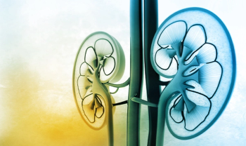
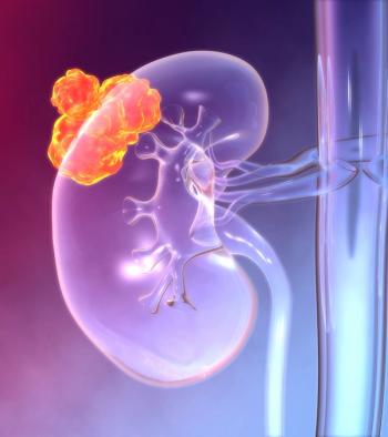
In patients with non–clear cell renal cell carcinoma (RCC), PD-L1 positivity is associated with worse clinical outcomes, including a shorter overall survival and time to recurrence, according to the results of a study recently
“This is the first study to shed light on the role of the PD-1/PD-L1 axis and PD-L1 expression in a group of tumors of the kidney that are uncommon and have no particular standard in the metastatic setting,” said Toni K. Choueiri, MD, of Dana-Farber Cancer Institute in Boston. “Like in clear cell RCC, PD-L1 expression (by immunohistochemistry) seemed to confer a worse outcome.”
According to Choueiri, with all the novel immunotherapies and immune checkpoint inhibitors being tested in clinic and with PD-L1 being a potential promising biomarker for this class of agents, the researchers thought to investigate the role of this biomarker in non–clear cell RCC. This histologic subtype represents 10% to 15% of all RCCs, and patients with this histology usually have been excluded from clinical trials with agents that target the PD-1 and PD-L1 axis.
In the study, the researchers obtained formalin-fixed paraffin-embedded specimens from 101 patients with non–clear cell RCC. Using immunohistochemistry in both the tumor cell membrane and tumor-infiltrating mononuclear cells, they evaluated for PD-L1 expression.
“We found that patients with non–clear cell RCC could express PD-L1 and that different types of non–clear cell RCCs have different levels of expression,” Choueiri told Cancer Network. “Furthermore, expression depends also on the scoring system, which ranges between 11% to 56%, depending on tumor cell membrane vs tumor-infiltrating mononuclear cells.”
Of the 101 patient samples, 10.9% were considered to be positive for PD-L1 when evaluated by tumor cells, including 5.6% of chromophobe RCC, 10% of papillary RCC, 30% of Xp11.2 translocation RCC, and 20% of collecting duct carcinoma. The results showed a significant association between PD-L1 positivity and higher cancer stage (P = .01) and grade (P = .03), as well as a shorter overall survival (P < .001).
In contrast, when evaluating PD-L1 positivity by the tumor-infiltrating mononuclear cells, 56.4% of the patient samples were positive for PD-L1, including 36.1% of chromophobe RCC, 60% of papillary RCC, 90% of Xp11.2 translocation RCC, and 100% of collecting duct carcinoma. Using this evaluation, there was only a trend toward shorter overall survival found in patients who were PD-L1–positive.
Both methods showed an association between PD-L1 positivity and a shorter time to recurrence.
“We found that patients with non–clear cell RCC do express the biomarker of interest for the class of agents that target PD-1 and PD-L1, and also that it seems that the expression is overall associated with poor prognosis,” Choueiri said. “We also feel that excluding these patients from trials of PD-1 inhibitors may be not fully justified if the reason for it is that these tumors lack the marker expression.”
Newsletter
Stay up to date on recent advances in the multidisciplinary approach to cancer.
Advertisement
Related Content
Advertisement

Latest CME


Advertisement
Advertisement
Trending on CancerNetwork
1
Modifiable Risk Factors Suggest Potential for Improving Cancer Prevention
2
2026 Tandem Meetings: What’s the Latest Research in Multiple Myeloma?
3
Dato-DXd Receives Priority Review in Unresectable/Metastatic TNBC
4
Barriers to CAR T-Cell Referral and Center Access in Multiple Myeloma
5