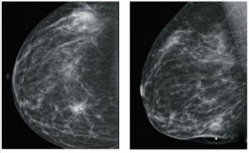
A 35-year-old woman noticed a mass in her right breast and underwent a diagnostic workup, including a mammogram that revealed a 2.4-cm mass and ultrasound that showed two adjacent masses, as well as enlarged axillary lymph nodes.

Your AI-Trained Oncology Knowledge Connection!


A 35-year-old woman noticed a mass in her right breast and underwent a diagnostic workup, including a mammogram that revealed a 2.4-cm mass and ultrasound that showed two adjacent masses, as well as enlarged axillary lymph nodes.
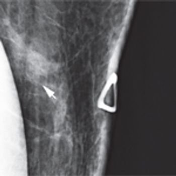
A 46-year-old woman had a routine screening mammogram that showed new calcifications in the posterior left breast. A diagnostic mammogram showed several small punctate calcifications, and a 6-month interval follow-up was recommended.

We review available strategies for screening and risk reduction through chemoprevention or risk-reducing surgery, as well as challenges for management of breast cancer in patients with prior exposure to radiation for Hodgkin lymphoma.
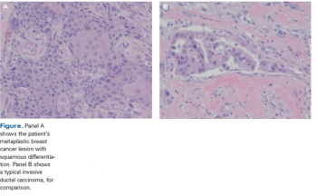
A 40-year-old woman noted a large mass in her right breast. A diagnostic mammogram and ultrasound confirmed a 3.4-cm mass with associated microcalcifications.
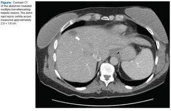
An asymptomatic 45-year-old woman presented for a screening mammogram and was noted to have a soft-tissue opacity with calcifications in the left breast. Ultrasound revealed a highly suspicious mass.
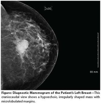
A 24-year-old woman presents to her primary care provider with a mass in her left breast. Examination confirms a 2.2-cm mass in the upper outer quadrant, with a single mobile axillary node that is firm to palpation.

Postpartum breast cancer represents a high-risk, under-recognized subset of young women’s breast cancer. The lack of clear identity for postpartum breast cancer is due in part to both the “dual effect” that pregnancy has on the incidence of breast cancer diagnosis.

A 40-year-old premenopausal woman with a new diagnosis of invasive lobular carcinoma occurring in a background of lobular carcinoma in situ presents to a multidisciplinary second opinion clinic.
This feature examines the case of a patient with newly diagnosed breast cancer in the setting of a first-trimester pregnancy presenting to our multidisciplinary breast cancer clinic.

patient is a 39-year-old premenopausal woman who presents with a new diagnosis of breast cancer to our multidisciplinary second opinion clinic.
The patient presented to her primary care physician 3 months prior with an inverted left nipple and a palpable lump that was highly suggestive of neoplasm on mammogram. An ultrasound-guided core biopsy revealed an infiltrating solid-type ductal carcinoma in situ. The estimated size of the mass was approximately 1 cm. She had no symptoms suggestive of metastatic disease.

Published: September 15th 2014 | Updated:

Published: April 15th 2011 | Updated:
Published: October 15th 2014 | Updated:
Published: September 15th 2015 | Updated:
Published: August 17th 2009 | Updated:

Published: September 1st 2007 | Updated: