- ONCOLOGY Vol 25 No 5
- Volume 25
- Issue 5
Chronic Inflammation and Cancer: The Role of the Mitochondria
We review the evidence implicating a strong association between chronic inflammation and cancer, with an emphasis on colorectal and lung cancer.
Accumulating evidence shows that chronic inflammation can promote all stages of tumorigenesis, including DNA damage, limitless replication, apoptosis evasion, sustained angiogenesis, self-sufficiency in growth signaling, insensitivity to anti-growth signaling, and tissue invasion/metastasis. Chronic inflammation is triggered by environmental (extrinsic) factors (eg, infection, tobacco, asbestos) and host mutations (intrinsic) factors (eg, Ras, Myc, p53). Extensive investigations over the past decade have uncovered many of the important mechanistic pathways underlying cancer-related inflammation. However, the precise molecular mechanisms involved and the interconnecting crosstalk between pathways remain incompletely understood. We review the evidence implicating a strong association between chronic inflammation and cancer, with an emphasis on colorectal and lung cancer. We summarize the current knowledge of the important molecular and cellular pathways linking chronic inflammation to tumorigenesis. Specifically, we focus on the role of the mitochondria in coordinating life- and death-signaling pathways crucial in cancer- related inflammation. Activation of Ras, Myc, and p53 cause mitochondrial dysfunction, resulting in mitochondrial reactive oxygen species (ROS) production and downstream signaling (eg, NFκB, STAT3, etc.) that promote inflammation- associated cancer. A recent murine transgenic study established that mitochondrial metabolism and ROS production are necessary for K-Ras–induced tumorigenicity. Collectively, inflammation-associated cancers resulting from signaling pathways coordinated at the mitochondrial level are being identified that may prove useful for developing innovative strategies for both cancer prevention and cancer treatment.
Introduction
Virchow is credited with suggesting the causal link between inflammation and cancer in the 19th century.[1] He based his conclusion on the astute observation that tumors often developed in the setting of chronic inflammation and that inflammatory cells were present in tumor biopsy specimens. Accumulating evidence that has emerged in the last decade or so has shed light on the underlying mechanisms accounting for the strong association between chronic inflammation and each step of tumorigenesis.[reviewed in 2-8] Notably, nearly 90% of all cancers are due to environmental factors and somatic mutations, whereas causal germ-line mutations are infrequent.[6] Nearly 20% of cancer deaths worldwide are attributed to chronic infection and/or inflammation, with gastrointestinal and lung cancers accounting for a substantial portion of the total burden.[1,9] An estimated 30% of cancers may be linked to exposure to tobacco and/or other airborne pollutants, and 20% can be attributed to chronic infections.[9] In general, a normal adaptive immune response is anti-tumorigenic; however, dysregulated innate and/or adaptive immune responses can be pro-tumorigenic. Human neutrophils can induce malignant transformation, which suggests that phagocytic cells are carcinogenic.[10] Mantovani et al[3,4] proposed that genetic instability resulting from cancer-related inflammation represents the seventh hallmark of tumorigenesis, in addition to the six proposed by Hanahan and Weinberg[11] (limitless replication, sustained angiogenesis, evasion of apoptosis, self-sufficiency in growth signals, insensitivity to anti-growth signals, and tissue invasion/metastasis).
In this review, we summarize the current knowledge supporting the association between chronic inflammation and cancer, highlighting the information that has been published since our 2002 ONCOLOGY review.[2] We then review the emerging evidence regarding important molecular and cellular pathways that link chronic inflammation to cancer. Emphasis will be placed on the pivotal role of the mitochondria in coordinating life- and death-signaling pathways important in inflammation-associated cancer. Collectively, the studies we review are revealing the crucial mechanisms that underlie inflammation-associated cancer and that may prove useful for developing novel cancer preventative and therapeutic strategies.
TABLE 1
Cancers Associated With Chronic Inflammation
Cancers Associated With Chronic Inflammation
Epidemiological evidence firmly supports a link between chronic inflammation and cancer that occurs in various organs (
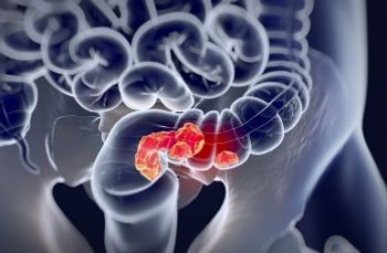
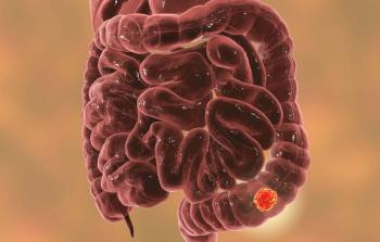
The best-established link between chronic inflammation and cancer is seen in colorectal cancer that develops in patients with inflammatory bowel disease (IBD; eg, ulcerative colitis and Crohn disease). These patients have a five- to seven-fold increased risk of developing colorectal cancer.[12-15] Nearly 43% of patients with ulcerative colitis develop colorectal cancer after 25 to 35 years.[15] Therapeutic strategies for the treatment or prevention of IBD aim to reduce the endogenous levels of tumor necrosis factor (TNF)-α, which is a key pathophysiologic element of the disease.[16] NFκB regulates multiple pathways involved in inflammation-associated cancer (eg, cytokine expression, angiogenesis, apoptosis, and COX-2 expression). TNF-α regulates NFκB, in part by receptor-mediated activation of inhibitory κB kinases (IKK) that stimulate degradation of proteins responsible for retaining the transcription factor in the cytosol, thereby enabling the translocation of NFκB to the nucleus. In a murine model of IBD, the development of colitis-associated colorectal cancer can be inhibited either by blocking TNF-α expression or by generating mice with colon epithelial cells that are deficient in IKK-β.[16,17] These findings in mice concur with the clinical observation that inhibition of the NFκB-regulated protein COX-2 by nonsteroidal anti-inflammatory drugs (NSAIDs) reduces the risk for colorectal cancer in humans with IBD by nearly 80%.[18,19] The synthesis of prostaglandin E2 (PGE2) by COX-2 induces the production of inflammatory cytokines such as interleukin (IL)-6.[20] Exposure to inflammatory cytokines (eg, IL-6, IL-10) causes the activation of the signal-transducing STAT proteins that work in conjunction with NFκB to regulate many genes involved in tumorigenesis.[reviewed in 4,21-23] The importance of STAT3 in colorectal cancer is evident in the finding that the development of tumors in a murine model of IBD is reduced in STAT3-deficient mice and through pharmacologic inhibition of IL-6.[22] The STAT pathway also regulates erythropoiesis and angiogenesis, both of which augment the availability of oxygenated blood to otherwise hypoxic tumors.[4,21-23] This pathway would provide an indirect mechanism for STAT-mediated tumor promotion. Collectively, these investigations provide the molecular basis for future studies on the role of inflammatory signaling through TNF-α, NFκB, STAT3, IL-6, and other signaling proteins in the etiology of inflammation-associated cancer. Hopefully, the information gained will prove useful in the management of colorectal cancer as well as IBD.
Lung cancer causes nearly 1 million deaths worldwide every year and is the leading cause of cancer deaths.[24] Although tobacco exposure is evident in nearly 90% of all patients with lung cancer, other chronic airway inflammatory conditions (eg, asbestosis, silicosis, exposure to airborne particulate matter (PM), idiopathic pulmonary fibrosis, tuberculosis, etc) are all independent risk factors for lung cancer and may account for a proportion of the non-smoking related cases.[25] Tobacco smoke contains nearly 5000 reactive chemicals, including over 1015 free radicals in the gas phase and 1018 free radicals per gram in the tar phase.[25] These include H2O2, •OH, and organic radicals.[25] As reviewed in detail elsewhere,[26-28] chronic inflammation has a pivotal role in the pathogenesis of chronic obstructive pulmonary disease (COPD). Smokers with COPD have a 1.3- to 6-fold increased risk of lung cancer compared with smokers without COPD, and this is likely due to persistent lung inflammation.[2,27,29] A meta-analysis demonstrated a strong indirect relationship between forced expiratory volume in 1 second (FEV1) and lung cancer risk.[30] Low-grade emphysema, without airway obstruction, is an independent risk factor for the development of lung cancer.[31] Although beyond the scope of this review, some of the potentially important molecular mechanisms underlying cancer associated with tobacco-induced inflammation include the production of ROS, inflammatory signaling (eg, via TNF-α, NFκB, IL-6, and others), single nucleotide polymorphisms in inflammatory cytokines (IL-1α and IL-1β), and increased ceramide and epithelial growth factor receptor (EGFR) signaling.[26-28] Interestingly, COPD-like inflammation induced by nontypeable Haemophilus influenza, which is the most common bacteria colonizing the airways of patients with COPD, promotes K-Ras–induced lung cancer in mice.[32] Notably, a recent study showed that mitochondrial metabolism is crucial for allowing mitochondrial ROS production at the Qo site of complex III, and that mitochondrial metabolism and ROS production were both required for mediating K-Ras–induced lung cancer in mice.[33] Macrophage migration inhibitory factor, an inflammatory cytokine, is produced at sites of bleomycin-induced lung injury in mice and functions to prevent apoptosis and promote tumor growth.[34] These innovative studies reveal insights into the pathogenesis of lung cancer occurring in the setting of emphysema-associated inflammation and should provide a rationale for future novel treatment strategies. Additional studies are necessary to understand why inflammation persists after smoking cessation as well as how inflammation in patients with COPD modulates disease expression.[29,35]
Lung cancer can also result from chronic pulmonary inflammation and fibrosis following exposure to other environmental toxins (eg, asbestos, silica, PM, beryllium). Further, a large cohort analysis of data from the Prostate, Lung, Colorectal and Ovarian Cancer Screening Trial showed that pulmonary scarring was associated with an elevated lung cancer risk (hazard ratio 1.5; 95% confidence interval 1.2-1.8).[36] In this section we highlight the role of asbestos. Asbestos is a term for a group of naturally occurring hydrated silicate fibers whose resilient strength and chemical properties make them ideal for a variety of building and insulation purposes. Asbestos causes an estimated 100,000 to 140,000 lung cancer deaths per year worldwide and contributes to nearly 5% to 7% of all lung cancers.[37,38] There are two classes of asbestos fibers: (1) serpentine fibers-curly-stranded structures, among which chrysotile is the principal commercial variety, and (2) amphibole fibers-straight, rod-like fibers (eg, crocidolite, amosite, tremolite, and others). Compared to chrysotile, amphibole fibers are more fibrogenic and carcinogenic, in part because their biopersistence in the lung results in chronic inflammation. Asbestos is an established carcinogenic agent that can induce chronic inflammation of the lung and pleura, ROS production, DNA damage, and cell death in all the major lung target cells (eg, bronchial and alveolar epithelial cells [AEC], mesothelial cells).[see for review: 39,40] Substantial investigations have shown that the extent of AEC injury and lack of sufficient AEC repair are important determinants of pulmonary inflammation and fibrosis following exposure to a wide variety of noxious agents, including asbestos.[40] There is a direct correlation between the levels of asbestosis seen in asbestos workers and the risk of developing lung cancer.[41] Asbestos-induced ROS cause DNA damage, such as single- or double-strand breaks, intra- and inter-strand cross-linking, and base damage.[see 42,43 for reviews] Repair of these lesions in most instances will restore the physiologic DNA structure, but abnormal DNA repair may result in gene mutations, chromosomal aberrations, and ultimately cell transformation. Early studies in our group showed that the repair of complex, inflammation-associated DNA damage, such as that caused by the exposure of cells to activated neutrophils, is slow compared to the repair of single-strand breaks, suggesting that residual DNA damage may lead to mutations or other cellular abnormalities that can promote tumorigenesis.[44] ROS-induced DNA damage is implicated in mediating the synergistic effect between asbestos and cigarette smoke for lung cancer risk.[see for review: 45,46] Convincing evidence, reviewed elsewhere,[40] has established that asbestos induces AEC apoptosis via the mitochondria-regulated (intrinsic) death pathway and involves mitochondrial ROS production. Interestingly, studies in transgenic mice suggest that Rac1-mediated mitochondrial H2O2 production from asbestos-exposed alveolar macrophages is necessary for the induction of pulmonary fibrosis.[47] However, further studies are required to better understand the molecular mechanisms underlying the link between asbestos-induced inflammation/pulmonary fibrosis and lung cancer.
One possibility is that diverse environmental stimuli, including asbestos and other lung carcinogens (eg, silica), but not inert particulates, cause pulmonary inflammation and fibrosis via activation of Nalp3 inflammasomes, which can stimulate caspase-1.[48,49] Nalp3 is a member of the NLR family of over 20 proteins. These proteins contain multiple functional domains, including an N-terminal protein-protein interaction domain that is necessary for caspase activation, a caspase recruitment domain (CARD), a central nucleotide-binding domain, and a C-terminal leucine-rich repeat domain.[50] Nalp3 inflammasome formation occurs when activated Nalp3 recruits caspase-1 and ASC, an adaptor molecule, via CARD-CARD interactions. Asbestos- and silica-induced lung inflammatory cell recruitment, cytokine production (eg, of IL-1β and others), and silicosis are all reduced in mice deficient in Nalp3, ASC, or caspase-1.[48,49] Moreover, by using specific pharmacologic inhibitors and targeted murine knockouts, it was found that the factors that appear essential for Nalp3 inflammasome activation include fiber uptake into phagocytic cells, an intact actin cytoskeleton, and ROS generated by nicotinamide adenine dinucleotide phosphate (NADPH) oxidase during phagocytosis. Thus, asbestos- and silica-induced Nalp3 inflammasome activation may be a novel therapeutic target for treatment/prevention of the underlying causes of inflammation-associated cancer.
Mechanisms Underlying Inflammation-Associated Cancer
TABLE 2
Inflammation-Associated Cancer: Facts & Questions
The last decade has witnessed much insight into inflammation-associated cancer; however, major gaps in our understanding remain.
FIGURE
Molecular Mechanisms Involved in Inflammation-Related Cancer
Inflammatory Cells in Tumorigenesis
Although a wide variety of cancers are associated with chronic inflammation and/or infection (see
As reviewed in detail elsewhere[7,14,56], one of the most compelling arguments linking inflammation-associated cancer to tumorigenesis is the observation that drugs that inhibit the production of prostaglandins during inflammation reduce the risk of various cancers, such as colorectal, esophageal, gastric, lung, breast, and ovarian cancer. These drugs include nonspecific NSAIDs, such as aspirin, and selective COX-2 inhibitors. COX-2 is an inducible form of cyclo-oxygenase that is activated in chronic inflammation. It is highly expressed in nearly all tumors.[7] COX-2 expression is necessary and sufficient to induce tumorigenesis in multiple in vitro and animal models.[reviewed in 2,7] It mediates the production of certain inflammatory cytokines that can act as tumor promoters, such as IL-6.[57] Randomized clinical trials show that NSAIDs decrease colon adenoma formation, an important precursor of colorectal cancer.[7,56] In breast cancer cells, COX-2 overexpression induces oxidative stress as well as chromosomal abnormalities (eg, fusions, breaks, and tetraploidy) that contribute to tumorigenesis.[58] Despite these remarkable advances in our understanding, no anti-inflammatory strategy is currently approved to prevent or treat cancer, although several are under development (eg, anti–IL-6 therapy for multiple myeloma). As reviewed in detail elsewhere[3,59], additional studies are required to determine which patient populations are appropriate for cancer preventative agents that target COX-2 or other relevant signaling pathways.
The tumor microenvironment contains a wide variety of inflammatory and immune cells, cytokines, and chemokines that have pro- and anti-tumorigenic activity, the balance of which likely dictates clinical outcome.[2-8] Experimental in vivo evidence unequivocally establishing the role of particular immune/inflammatory cells and cytokines/chemokines in tumorigenesis is lacking.[6] The most common immune cells in tumors are tumor-associated macrophages (TAMs) and T cells. TAMs, which are the major source of cytokine production in the tumor microenvironment, promote tumorigenesis in several ways. They produce protein factors that stimulate tumor cell growth, directly and indirectly (eg, by stimulating angiogenesis), and they stimulate metastasis by producing matrix-degrading enzymes.[5,6] TAMs are classified either as M1 or M2 macrophages, depending on their response to various stimuli. M1 TAMs respond to interferon (IFN)-γ or microbial exposure by expressing high levels of cytokines involved in anti-tumor and anti-microbial activity (eg, TNF-α, IL-1, IL-6, IL-12, IL-23), while M2 TAMs are proangiogenic/tissue-remodeling macrophages that display reduced expression of IL-12 and increased expression of the anti-inflammatory cytokine IL-10 following exposure to IL-4, IL-10, or IL-13.[3,6] The M1 and M2 TAM phenotypes are plastic, based on their gene expression profiles.[6] The protumorigenic effects of TAMs are suggested by the finding that TNF-α–deficient mice are protected against drug-induced skin cancer.[60,61] Also, TAMs augment Wnt signaling via a TNF-α–dependent pathway in gastric cancer; this pathway is necessary for growth and for epithelial-mesenchymal cell transition that is important in metastasis.[62] Phase 1 and II clinical trials are underway examining the role of TNF-α antagonists in patients with renal cancer[63] as well as advanced cancers.[64] As reviewed in detail elsewhere,[5] studies in transgenic mice have established a protumorigenic role for IL-1. The finding of increased skin and colitis-related cancers occurring in mice deficient in the atypical chemokine receptor D6 establishes a prominent role for CC chemokines in tumorigenesis.[65] In this context, it is not surprising that a high tumor TAM content generally foreshadows a poor prognosis.[66]
T cells can also impact cancer outcomes. Increased levels of CD8+ cytotoxic T lymphocytes and CD4+ helper 1 (Th1) cells portend a better prognosis in certain tumors (eg, colon, melanoma, pancreatic, multiple myeloma, lung) and comprise a therapeutic approach to the treatment of these cancers.[6] In contrast, a T-cell deficiency can augment tumor formation.[6] Additional investigation is necessary to determine why certain T-cell subsets are pro-tumorigenic in one cancer but anti-tumorigenic in another. Also, it is unknown whether there is a common upstream inflammatory signal (eg, mitochondrial ROS production) that is activated in all malignancies, and if so, whether this regulates the balance between TAM and T-cell pro- and/or anti-tumorigenic activities.
Inflammation and Oncogenes/Tumor Suppressor Genes
Similar to Ras, the Myc oncogenes are mutated in many human cancers and alter mitochondrial function (eg, increased electron transport, oxygen uptake, and ROS production) in a way that induces the rapid cell growth that is a crucial element of tumorigenesis.[51] Growth factors and chemokines produced in the setting of inflammation-associated cancer augment Myc overexpression in cancer cells, thereby driving Ras activation and abnormal DNA synthesis.[4] In a murine Myc model of pancreatic cancer, the initial wave of angiogenesis is mediated by the inflammatory cytokine IL-1β.[69] Interestingly, a recent gene expression profile study showed that the Myc network of transcription programs accounts for most of the similarity between embryonic stem cells and cancer cells.[70] Myc activation can trigger mitochondria-regulated apoptosis, whereas Myc-induced DNA damage and cellular transformation are prevented by mitochondria-targeted antioxidants.[71,72] Thus, the emerging evidence suggests that the mitochondria are important downstream effector organelles in both Myc- and Ras-induced oncogenic transformation (see
The tumor suppressor protein p53 is an important transcriptional factor for multiple proteins involved in the cellular DNA damage response, and it is likely important in inflammation-associated cancer.[see for review 73] Following DNA damage caused by oxidative stress (eg, that resulting from exposure to tobacco, asbestos, etc), an intact p53 response prevents mutations from accumulating by increasing the expression of genes that inhibit cell growth, thereby increasing the time available for DNA repair. However, if DNA damage is extensive, p53 activation can augment apoptosis by inducing pro-apoptotic genes while inhibiting expression of anti-apoptotic genes, ultimately causing mitochondrial dysfunction and intrinsic apoptosis. Because of its central role in directing cellular life and death outcomes, it is not surprising that mutations in p53 gene family members are common in human tumors.[73] Mitochondrial ROS block wild-type p53 function and promote the formation of p53 mutations.[reviewed in 4,51] Mutations in p53, some from inflammation-associated oxidative stress, are evident in the epithelium of cancer cells and in inflamed, but non-dysplastic epithelial cells.[74] This suggests that genomic changes can result from chronic inflammation. Altered p53 expression has also been implicated in the pathophysiology of pulmonary fibrosis, including that due to asbestos, as well as in pulmonary fibrosis–associated bronchogenic lung cancer.[75-80] For example, increased p53 protein expression is detected in the bronchiolar and alveolar epithelium of humans with idiopathic pulmonary fibrosis and in rodents exposed to asbestos.[75-80]Furthermore, increased p53 levels are detected in lung cancers of patients with asbestosis,[81] and p53 point mutations are widely evident in the respiratory epithelium of smokers and asbestos-exposed individuals.[82] p53 mediates asbestos-induced, mitochondria-regulated apoptosis in lung epithelial cells, and this is blocked in cells incapable of producing mitochondrial ROS.[80] Notably, loss of p53 results in mtDNA depletion, altered mitochondrial function, and increased H2O2 production.[83] Considerable evidence, reviewed in detail elsewhere,[51] has established that p53 is a crucial regulator of mitochondrial function, including ROS generation and mtDNA repair following oxidative damage, as well as mitochondrial biogenesis and mtDNA replication. Although formal evidence is lacking, it is likely that loss of wild-type p53 function augments the deleterious effects induced by Ras and Myc on mitochondrial function described above.[51] Thus, p53 has a key role in regulating the response to cellular DNA damage caused by exposure to oxidative stress, and likely plays a role in the pathogenesis of inflammation-associated cancer. Future investigations are required to better understand how the Ras, Myc, and p53 pathways are interconnected.
As reviewed in detail elsewhere,[4,8] chronic inflammation can effect each of the six hallmarks of tumorigenesis identified by Hanahan and Weinberg,[11] including limitless replicative potential, sustained angiogenesis, evasion of apoptosis, self-sufficiency in growth signaling, insensitivity to anti-growth signals, and tissue invasion/metastasis. The evasion of immune surveillance mechanisms and genetic instability due to inflammation-associated cancer have each been proposed as the seventh hallmark of cancer-again emphasizing the role of inflammation in cancer.[4,84] Inflammation-associated cancer induces oxidative stress that can lead to DNA damage and cellular stress, which in turn cause abnormalities in mitosis (eg, through chromosomal abnormalities) and metabolism (eg, through the Warburg effect, or increased glucose uptake for glycolysis).[8] Although beyond the scope of this review, nearly 30 different cancer therapies targeting these assorted mechanistic hallmarks of inflammation-associated cancer are in various stages of development.[reviewed in 8] A crucial unresolved issue is whether inflammatory signaling in susceptible tissues (eg, the lungs of smokers) can be altered so as to favor adaptive immunity (anti-tumorigenic activity) rather than pro-tumorigenic activity.
Inflammation, ROS, and the Mitochondria
α, IL-1β, NFκB, STAT3, and COX-2) decreases the incidence and spread of certain tumors (eg, colorectal cancer). In general, inflammatory /immune components necessary at one stage of tumorigenesis may be completely dispensable during another stage.[3,6] Also, adaptive transfer of inflammatory cells or overexpression of certain cytokines promotes tumor formation.[3]
The major sources of ROS in the setting of inflammation-associated cancer include (1) NADPH oxidase present in phagocytes and other cells and (2) mitochondria. In non-phagocytic cells, over 95% of ROS formed during normal metabolism originate from the electron transport chain (ETC) in the inner mitochondrial membrane in close proximity to mtDNA.[51] ROS-induced mtDNA damage is implicated in a wide range of pathologic processes, including carcinogenesis, aging, and degenerative diseases.[85,86] Emerging studies suggest that mitochondrial ROS form crucial intermediates between environmental and host stimuli that result in inflammation-associated cancer (see
Given the close proximity of mtDNA to the mitochondrial ETC and the lack of protective histones, mtDNA damage resulting from oxidative stress may be important in the pathogenesis of inflammation-associated cancer. For example, with asbestos, lung mesothelial cell mtDNA damage is evident following exposure to a four-fold lower concentration of crocidolite asbestos than the crocidolite doses required to cause nuclear DNA damage.[90] Also, several lines of evidence implicate mtDNA oxidative injury as a key trigger of apoptosis that may be important in inflammation-associated cancer, including: (1) that cell death is more closely associated with mtDNA oxidative lesions than with nuclear DNA lesions, (2) that mtDNA damage precedes ATP depletion and mitochondrial dysfunction, (3) that enhancing mtDNA repair blocks cell death, and (4) that deficiency of mtDNA repair enhances cell death.[reviewed in 51,86] Base excision repair (BER) is the principal pathway for repairing oxidative mtDNA damage.[83] Epidemiological data suggest that the levels of 8OHdG, the most common DNA base change arising from oxidative stress, is linked with various cancers and neurodegenerative diseases.[85,86,91-94] 8OHdG induces mutations in replicating cells by preferentially mispairing with adenine during DNA synthesis, thereby increasing the incidence of G:C to T:A transversions. DNA glycosylases have a key role in BER pathways: they recognize the oxidized DNA adduct and excise the damaged base. 8-oxo-guanine DNA glycosylase (Ogg1), which is responsible for repairing 8OHdG, has a dual function: it preferentially recognizes 8OHdG opposite cytosine and then excises it via its apurinic/apyrimidic lyase activity. All mtDNA BER repair proteins, including Ogg1, are nuclear-encoded and imported into mitochondria.[83] Overexpression of mitochondria-targeted Ogg1 blocks intrinsic apoptosis in ROS-exposed vascular endothelial and asbestos-exposed HeLa cells.[90,95,96] We recently extended these findings to AEC exposed to oxidative stress (asbestos or H2O2).[97] Further, using Ogg1 mutants incapable of 8OHdG DNA repair, we showed that Ogg1 functions in a role independent of DNA repair by preserving mitochondrial aconitase levels. Mitochondrial aconitase has a dual role: (1) it serves as an iron-sulfur– containing tricarboxycylic acid cycle enzyme that is a mitochondrial redox-sensor susceptible to oxidative degradation and (2) it maintains mtDNA by mechanisms that are independent of its catalytic activity.[98-100] Mitochondrial aconitase co-precipitates with frataxin, an iron chaperone protein that is as good as Ogg1 at preventing aconitase oxidative inactivation.[97,101] Given the importance of p53 in inflammation-associated cancer, it is of interest that Ogg1 is under transcriptional regulation by p53.[102,103] Collectively, these findings suggest critical crosstalk between the mitochondria (ROS, aconitase, Ogg1, etc) and p53 that is likely important in inflammation-associated cancer.
Activation of oncogenic transcription factors can be triggered through pattern recognition receptors, by exposure to components of bacteria, viruses, and interestingly, mtDNA.[104,105] Chronic inflammation /infection can lead to extensive cellular damage in target organs (eg, necrotic epithelial cells and macrophages in tumors), and this results in the release of damage-associated molecular pattern (DAMP) or pathogen-associated molecular pattern (PAMP) molecules.[reviewed in 6,56] DAMPs include IL-1α, high mobility group B1 molecule (HMGB1), and other molecules that work in concert to facilitate inflammation.[59] The underlying mechanisms are the subject of ongoing studies. Circulating mtDNA and mitochondrial DAMPs can be detected in patients with trauma, a finding that may account for the increased risk of multi-organ dysfunction in these patients.[105] These investigations illustrate the diverse mechanisms by which alterations in the mitochondria can impact inflammation-associated cancer. It is unclear whether epithelial cells or immune/inflammatory cells are the primary source of DAMPs in tumors. It will be of interest to determine whether chronic inflammation/tissue injury results in the release of mtDNA, and if so, whether this is crucial for driving inflammation-associated cancer. Further studies are necessary to better understand the precise molecular details by which mitochondrial respiration, mitochondrial ROS production, and mtDNA damage affect specific components of inflammation-associated cancer.
Inflammation and Tumor-Promoting Signaling Pathways
Tumor cells, carcinogen-exposed epithelial cells, and inflammatory cells utilize NFκB, a tightly regulated transcription factor, to activate a number of genes coding for proteins involved in inflammation-associated cancer, including cytokines, growth factors, adhesion molecules, angiogenic factors, proto-oncogenes (eg, Myc), COX-2, and nitric oxide synthase.[reviewed in: 3,6,7,106] NFκB, a dimer of two Rel-family proteins (p50 and p65), is activated in the cytoplasm by diverse cellular conditions including excess ROS, hypoxia, and HIF-1α. It is also regulated autonomously by genetic alterations that lead to phosphorylation of its inhibitor protein (IκBα). The phosphorylation of IκBα results in the proteolytic degradation and subsequent translocation of IκB to the nucleus, where it binds to and regulates the DNA.[106] NFκB is also activated downstream of signaling by inflammatory cytokines (eg, TNF-α, IL-1β) as well as by the toll-like receptor–MyD88 pathway that is stimulated by microbes and tissue damage.[4] NFκB can have divergent effects in various models of carcinogenesis that likely relate to the balance between activating downstream pro- and anti-tumorigenic effects.[106-109)] Murine transgenic studies have established a key role for NFκB signaling pathways in colitis-associated cancer, liver cancer, and breast cancer metastasis.[reviewed in 4,7,14] NFκB activation by TNF-α augments nuclear entry of Wnt/β-catenin in inflammation-associated gastric cancer,[110] as well as in colonic crypt cells[111]-a finding that is likely crucial for promoting tissue invasion/metastasis. Asbestos causes prolonged, dose-dependent transcriptional activation of NFκB-dependent genes in vitro and in vivo by a ROS-dependent mechanism.[reviewed in 112] In murine models that inhibit IKKβ-dependent NFκB activation, acute inflammation is exacerbated while chronic intestinal inflammation is attenuated.[113] These findings underscore how critical the context of inflammation (eg, acute vs chronic) is in regulating the pro-inflammatory and anti-apoptotic effects of NFκB. The collective evidence suggests that NFκB has primarily pro-tumorigenic effects but that an anti-inflammatory role can occur. Further studies are necessary to determine the precise role of pharmacologic and genetic targeting of the NFκB-dependent pathways in various cancer preventative and treatment strategies.
STAT3, like NFκB, is a transcription factor that is often constitutively activated in tumors and immune cells. It mediates a number of crucial tumorigenic signaling pathways (eg, cell proliferation, apoptosis, Myc expression, evasion of immune surveillance).[reviewed in 4] The STAT family contains seven members, but STAT3 has been most closely implicated in inflammation-associated cancer.[reviewed in 21] STAT3 signaling is essential for stem-cell renewal as well as for persistent NFκB activation in tumor cells.[114,115] Further, mitochondrial STAT3 is essential for Ras-dependent oncogenic transformation.[116] The molecular mechanism(s) that account for the presence of STAT3 in the mitochondria are unclear, but apparently do not depend on increased STAT3 transcriptional activity, nor on changes in mtDNA-encoded proteins. Rather, the presence of mitochondrial STAT3 appears to be mediated by greater mitochondrial ETC activity. A firm role for STAT3 in colitis-associated cancer is suggested by the finding of a reduced incidence of colon cancers in STAT3-deficient mice.[reviewed in 14] Also, a colitis-inducing strain of Bacteroides fragilis that is implicated in colorectal cancer is a potent activator of STAT3 in humans and mice.[117] Mutations in EGFR result in downstream IL-6 production and STAT3 phosphorylation in lung adenocarcinomas.[118,119] Although the precise molecular details await further study, the available experimental evidence supports an important role for the interconnected signaling cascade of NFκB–IL-6–STAT3 in the development of inflammation-associated cancer.
Conclusions
Cancer-related inflammation remains a significant challenge to healthcare providers, as well as to investigators studying the basic mechanisms underlying tumorigenesis. Largely because the pathogenesis of inflammation-associated cancer is incompletely understood, there are currently limited therapeutic techniques for modifying cancers that occur in the setting of chronic inflammation. The accumulating evidence links a wide variety of chronic inflammatory conditions to diverse groups of cancers (see
Financial Disclosure:The authors have no significant interest or other relationship with the manufacturers of any products or providers of any service mentioned in this article.
References:
REFERENCES:
1. Balkwill F, Mantovani A. Inflammation and cancer: back to Virchow? Lancet. 2001;357:539-45.
2. Shacter E, Weitzman SA. Chronic inflammation and cancer. Oncology. 2002;16:217-29.
3. Mantovani A, Allavena P, Sica A, Balkwill F. Cancer-related inflammation. Nature. 2008;454:436-44.
4. Colotta F, Allavena P, Sica A, Garlanda C, Mantovani A. Cancer-related inflammation, the seventh hallmark of cancer: links to genetic instability. Carcinogenesis. 2009;30:1073-81.
5. Mantovani A, Garlanda C, Allavavena P. Molecular pathways and targets in cancer-related inflammation. Ann Med. 2010;42:161-70.
6. Grivennikov SI, Greten FR, Karin M. Immunity, inflammation, and cancer. Cell. 2010;140:883-99.
7. Schetter AJ, Heegaard NHH, Harris CC. Inflammtion and cancer: interweaving microRNA, free radical, cytokine and p53 pathways. Carcinogenesis. 2010;31:37-49.
8. Lio J, Solimini NL, Elledge SJ. Principles of cancer therapy: oncogene and non-oncogene addiction. Cell. 2009;136:823-37.
9. Aggarwal BB, Vijayalesshmi RV, Sung B. Targeting inflammatory pathways for prevetion and therapy of cancer: short-term friend, long-term foe. Clin Cancer Res. 2009;15:425-30.
10. Weitzman SA, Weitberg AB, Clark E, Stossel TP. Phagocytes as carcinogens: malignant transformation produced by human neutrophils. Science. 1985;227:
1231-3.
11. Hanahan D, Weinberg RA. The hallmarks of cancer. Cell. 2000;100:50-70.
12. Ekbom A, Helmick C, Zack M, Adami HO. Ulcerative colitis and colorectal cancer. A population-based study. N Engl J Med. 1990;323:1228-33.
13. Gillen CD, Walmsley RS, Prior P, et al. Ulcerative colitis and Crohn's disease: a comparison of the colorectal cancer risk in extensive colitis. Gut. 1994;35:1590-2.
14. Ferrone C, Dranoff G. Dual roles for immunity in gastrointestinal cancers. J Clin Oncol. 2010;28:4045-51.
15. Ekbom A. Risk of cancer in ulcerative colitis. J Gastrointest Surg. 1998;2:312-3.
16. Popivanova BK, Kitamura K, Wu Y, et al. Blocking TNF-alpha in mice reduces colorectal carcinogenesis associated with chronic colitis. J Clin Invest. 2008;118:560-70.
17. Greten FR, Eckmann L, Greten TF, et al. IKKbeta links inflammation and tumorigenesis in a mouse model of colitis-associated cancer. Cell. 2004;118:285-96.
18. Kopp E, Ghosh S. Inhibition of NF-κB by sodium salicylate and aspirin. Science. 1994;265:956-9.
19. Eaden J, Abrams K, Ekbom A, et al. Colorectal cancer prevention in ulcerative colitis: a case-control study. Aliment Pharmacol Ther. 2000;14:145-53.
20. Williams JA, Shacter E. Regulation of macrophage cytokine production by prostaglandin E2: Distinct roles of Cox-1 and Cox-2. J Biol Chem. 1997;41:25693-9.
21. Yu H, Pardoll D, Jove R. STATs in cancer inflammation and immunity: A leading role for STAT3. Nat Rev Cancer. 2009; 9:798-809.
22. Grivennikov S, Karin E, Terzic J, et al. IL-6 and Stat3 are required for survival of intestinal epithelial cells and development of colitis-associated cancer. Cancer Cell. 2009;15:103-13.
23. Kujawski M, Kortylewski M, Lee H, et al. Stat3 mediates myeloid cell-dependent tumor angiogenesis in mice. J Clin Invest. 2008;118:3367-77.
24. Sun S, Schiller JH, Gazdar AF. Lung cancer in never smokers – a different disease. Nat Rev Cancer 2007;7:778-90.
25. Alberg AJ, Samet JM. Epidemiology of lung cancer. Chest. 2003;123:21S-49S.
26. Goldkorn T, Filosto S. Lung injury and cancer. Am J Respir Cell Mol Biol. 2010;43:259-68.
27. Yao H, Rahman I. Current concepts on the role of inflammation in COPD and lung cancer. Curr Opinions Pharmacol. 2009;9:375-83.
28. Hogg JC, Timens W. The pathology of chronic obstructive pulmonary disease. Annu Rev Pathol. 2009;4:435-59.
29. Young RP, Hopkins RJ, Christmas T, et al. COPD prevalence is increased in lung cancer, independent of age, sex and smoking history. Eur Respir J. 2009;34:380-6.
30. Wasswa-Kintu S, Gan WQ, Man SF, et al. Relationship between reduced forced expiratory volume in one second and the risk of lung cancer: a systematic review and meta-analysis. Thorax. 2005;
60:570-5.
31. Caballero A, Torres-Duque CA, Jaramill C, et al. Prevalence of COPD in five Columbian cities situated at low, medium and high altitude (PREPOCOL study). Chest. 2008;133;343-9.
32. Moghaddam SJ, Li H, Cho S-N, et al. Promotion of lung carcinogenesis by chronic obstructive pulmonary disease-like airway inflammation in a K-ras-induced mouse model. Am J Respir Cell Mol Biol. 2009;40:443-53.
33. Weinberg F, Hammanaka R, Wheaton WW, et al. Mitochondrial metabolism and ROS generation are essential for Kras-mediated tumorigenicity. PNAS. 2010;107:8788-93.
34. Arenberg D, Luckhardt TR, Carskadon S, et al. Macrophage migration inhibitory factor promotes tumor growth in the context of lung injury and repair. Am J Respir Crit Care Med. 2010;182:1030-7.
35. Cosio MG, Seatta M, Agusti A. Immunologic aspects of chronic obstructive pulmonary disease. N Engl J Med. 2009;360:2445-54.
36. Yu YY, Pinsky PF, Caporaso NE, et al. Lung cancer risk following detection of pulmonary scarring by chest radiography in the Prostate, Lung, Colorectal, and Ovarian Cancer Screening Trial. Arch Intern Med. 2008;168:2326-32.
37. LaDou FJ. The asbestos cancer epidemic. Environ Health Perspect. 2004;112:285-90.
38. Tossavainen A. Global use of asbestos and the incidence of mesothelioma. Int J Occup Environ Health. 2004;10:22-5.
39. Heintz NH, Janssen-Heininger YM, Mossman BT. Asbestos, lung cancers, and mesotheliomas: from molecular approaches to targeting tumor survival pathways. Am J Respir Cell Mol Biol. 2010;42:133-9.
40. Liu G, Beri R, Mueller A, Kamp DW. Molecular mechanisms of asbestos-induced lung epithelial cell apoptosis. Chem-Biol Interactions. 2010;188:309-18.
41. Weiss W. Asbestosis: a marker for the increased risk of lung cancer among workers exposed to asbestos. Chest. 1999;115:536-49.
42. Jaurand MC, Mechanisms of fiber-induced genotoxicity, Environ Health Perspect. 1997;105(Suppl 5):1073-84.
43. Upadhyay D, Kamp DW. Asbestos-induced pulmonary toxicity: role of DNA damage and apoptosis, Exp Biol Med (Maywood). 2003;228:650-9.
44. Shacter E, Beecham EJ, Covey JM, et al. Activated neutrophils induce prolonged DNA damage in neighboring cells. Carcinogenesis. 1988; 9:2297-304
45. Kamp DW, Graceffa P, Pryor WA, Weitzman SA. The role of free radicals in asbestos-induced diseases. Free Radic Biol Med. 1992;12:293-315.
46. Nelson HH, Kelsey KT. The molecular epidemiology of asbestos and tobacco in lung cancer. Oncogene. 2002;21:7284-8.
47. Murthy S, Ryan A, He C, et al. Rac1-mediated mitochondrial H2O2 generation regulates MMP-9 gene expression in macrophages via inhibition of SP-1 and AP-1. J Biol Chem. 2010;285:25062-73.
48. Dostert C, Petrilli V, Van Bruggen R, et al. Innate immune activation through Nalp3 inflammasome sensing of asbestos and silica. Science. 2008;320:674-7.
49. Cassel SL, Eisenbarth SC, Iyer SS, et al. The Nalp3 inflammasome is essential for the development of silicosis. PNAS. 2008;105:935-40.
50. Petrilli V, Dostert C, Muruve DA, Tschopp J. The inflammasome: a danger sensing complex triggering innate immunity. Curr Opin Immunol. 2007;19:615-22.
51. Ralph SJ, Rodriguez-Enriquez S, Neuzil J, et al. The causes of cancer revisited: “mitochondrial malignancy” and ROS-induced oncogenic transformation – Why mitochondria are targets for cancer therapy. Mol Aspects Med. 2010;31:145-70.
52. Weinberg F, Chandel NS. Reactive oxygen species-dependent signaling regulates cancer. Cell Mol Life Sci. 2009;66:3663-73.
53. Sato Y, Takahashi S, Kinouchi Y, et al. IL-10 deficiency leads to somatic mutations in a model of IBD. Carcinogenesis. 2006;27:1068-73.
54. Gungor N, Godschalk RWL, Pachen DM, et al. Activated neutrophils inhibit nucleoside excision repair in human pulmonary epithelial cells: role of myeloperoxidase. FASEB. 2007;21:2359-67.
55. Rao VP, Poutahidis T, Ge Z, et al. Innate immune inflammatory response against enteric bacteria Helicobacter hepaticus induces mammary adenocarcinoma in mice. Cancer Res. 2006;66:7395-400.
56. Cuzick J, Otto F, Baron JA, et al. Aspirin and non-steroidal anti-inflammatory drugs for cancer prevention: an international consensus statement. Lancet Oncol. 2009;10:501-7.
57. Hinson RM, Williams JA, Shacter E. Elevated interleukin-6 is induced by prostaglandin E2 in a murine model of inflammation. Possible role of cyclooxygenase 2. Proc Natl Acad Sci USA. 2006;93:4885-90.
58. Singh B, Cook KR, Vincent L, et al. Cyclooxygenase-2 induces genomic instability, BCL2 expression, doxorubicin resistance, and altered cancer-initiating cell phenotype in MCF7 breast cancer cells. J Surg Res. 2008;147:240-6.
59. Demaria S, Pikarsky E, Karin M, et al. Cancer and inflammation: promise for biologic therapy.
J Immunother. 2010;33:335-51.
60. Moore RJ, Owens DM, Stamp G, et al. Mice deficient in tumor necrosis factor-alpha are resistant to skin carcinogenesis. Nat Med 1999;5:828-31.
61. Balkwill F. Tumor necrosis factor and cancer. Nat Rev Cancer. 2009;9:361-71.
62. Oguma K, Oshima H, Aoki M, et al. Activated macrophages promote Wnt signaling through tumor necrosis factor-alpha in gastric tumour cells. EMBO
J. 2008;27:1671-81.
63. Harrison ML, Obermueller E, Maisey NR, et al. Tumor necrosis factor alpha as a new target for renal cell carcinoma: two sequential phase II trials of infliximab at standard and high dose. J Clin Oncol. 2007;
25:4542-9.
64. Brown ER, Charles KA, Hoare SA, et al. A clinical study assessing the tolerability and biological effects of infliximab, a TNF-alpha inhibitor, in patients with advanced cancer. Ann Oncol. 2008;19:1340-6.
65. Nibbs RJ, Gilchrist DS, King V, et al. The atypical chemokine receptor D6 suppresses the development of chemically induced skin tumors. J Clin Invest. 2007;117:1884-92.
66. Murdoch C, Muthana M, Coffelt SB, Lewis CE. The role of myeloid cells in the promotion of tumour angiogenesis. Nat Rev Cancer. 2008;8:618-31.
67. Chin LJ, Ratner E, Leng S, et al. A SNP in a let-7 microRNA complementary site in the KRAS 3' untranslated region increases non-small cell lung cancer risk. Cancer Res. 2008;68:8535-40.
68. Christensen BC, Moyer BJ, Avissar M, et al. A let-7 microRNA-binding site polymorphism in the KRAS 3' UTR is associated with reduced survival in oral cancers. Carcinogenesis. 2009;30:1003-7.
69. Shchors K, Shchors E, Rostker F, et al. The Myc-dependent angiogenic switch in tumors is mediated by interleukin 1beta. Genes Dev. 2006;20:2527-38.
70. Kim J, Woo AJ, Chu J, et al. A Myc network accounts for similarities between embryonic stem and cancer cell transcription programs. Cell. 2010;143:313-24.
71. Morrish F, Neretti N, Sedivy JM, Hockenbery DM. The oncogene c-Myc coordinates regulation of metabolic networks to enable rapid cell cycle entry. Cell Cycle. 2008;7:1054-66.
72. K C S, Cárcamo JM, Golde DW. Antioxidants prevent oxidative DNA damage and cellular transformation elicited by the over-expression of c-MYC. Mutat Res. 2006;593:64-79.
73. Vousden KH, Prives C. Blinded by the light: the growing complexity of p53. Cell. 2009;137:413-31.
74. Kraus S, Arber N. Inflammation and colorectal cancer. Curr Opin Pharmacol. 2009;9:405-10.
75. Kuwano K, Kunitake R, Kawasaki M, et al. P21Waf1/Cip1/Sdi1 and p53 expression in association with DNA strand breaks in idiopathic pulmonary fibrosis. Am J Respir Crit Care Med. 1996;154:477-83.
76. Johnson NF, Jaramillo RJ. p53, Cip1, and Gadd153 expression following treatment of A549 cells with natural and man-made vitreous fibers. Environ Health Perspect. 1997;105(Suppl 5):1143-5.
77. Mishra A, Liu JY, Brody AR, Morris GF. Inhaled asbestos fibers induce p53 expression in the rat lung. Am J Respir Cell Mol Biol. 1997;16:479-85.
78. Burmeister B, Schwerdtle T, Poser I, et al. Effects of asbestos on initiation of DNA damage, induction of DNA-strand breaks, P53-expression and apoptosis in primary, SV40-transformed and malignant human mesothelial cells. Mutat Res. 2004;558:81-92.
79. Plataki M, Koutsopoulos AV, Darivianaki K, et al. Expression of apoptotic and antiapoptotic markers in epithelial cells in idiopathic pulmonary fibrosis. Chest. 2005;127:266-74.
80. Panduri V, Surapureddi S, Soberanes S, et al. P53 mediates amosite asbestos-induced alveolar epithelial cell mitochondria-regulated apoptosis. Am J Respir Cell Mol Biol. 2006;34:443-52.
81. Nuorva K, Makitaro R, Huhti E, et al. p53 protein accumulation in lung carcinomas of patients exposed to asbestos and tobacco smoke. Am J Respir Crit Care Med. 1994;150:528-33.
82. Husgafvel-Pursiainen K, Kannio A, Oksa P, et al. Mutations, tissue accumulations, and serum levels of p53 in patients with occupational cancers from asbestos and silica exposure. Environ Mol Mutagen. 1997;30:224-30.
83. Lebedeva MA, Eaton JS, Shadel GS. Loss of p53 causes mitochondrial DNA depletion and altered mitochondrial reactive oxygen species homeostasis. Biochim Biophys Acta. 2009;1787:328-34.
84. Kroemer G, Pouyssegur J. Tumor cell metabolism: cancer's Achilles' heel. Cancer Cell. 2008;13:472-82.
85. Bohr VA, Stevnsner T, de Souza-Pinto NC. Mitochondrial DNA repair of oxidative damage in mammalian cells. Gene. 2002;286:127-34.
86. Van Houten B, Woshner V, Santos JH. Role of mitochondrial DNA in toxic responses to oxidative stress. DNA Repair (Amst). 2006;5:145-52.
87. Tormos KV, Chandel NS. Inter-connection between mitochondria and HIFs. J Cell Mol Med. 2010;14:795-804.
88. Calore F, Genisset C, Casellato A, et al. Endosome-mitochondria juxtaposition during apoptosis induced by H. pylori VacA. Cell Death Differ. 2010;17:1707-16.
89. Rao VA, Klein SR, Bonar SJ, et al. The antioxidant transcription factor Nrf2 negatively regulates autophagy and growth arrest induced by the anticancer redox agent mitoquinine. J Biol Chem. 2010;
285:34447-59.
90. Shukla A, Jung M, Stern M, et al. Asbestos induces mitochondrial DNA damage and dysfunction linked to the development of apoptosis. Am J Physiol Lung Cell Mol Physiol. 2003;285:L1018-25.
91. Garrido C, Kroemer G. Life's smile, death's grin: vital functions of apoptosis-executing proteins. Curr Opin Cell Biol. 2004;16:639-46.
92. Russo MT, De Luca G, Degan P, et al. Accumulation of the oxidative base lesion 8-hydroxyguanine in DNA of tumor-prone mice defective in both the Myh and Ogg1 DNA glycosylases. Cancer Res. 2004;64:4411-14.
93. Ames BN, Shigenaga MK, Hagen TM. Oxidants, antioxidants, and the degenerative diseases of aging. Proc Natl Acad Sci U S A. 1993;90:7915-22.
94. Chevillard S, Radicella JP, Levalois C, et al. Mutations in OGG1, a gene involved in the repair of oxidative DNA damage, are found in human lung and kidney tumours. Oncogene. 1998;16:3083-6.
95. Dobson AW, Grishko V, LeDoux SP, et al. Enhanced mtDNA repair capacity protects pulmonary artery endothelial cells from oxidant-mediated death. Am J Physiol Lung Cell Mol Physiol. 2002;283:L205-10.
96. Rachek LI, Grishko VI, Ledoux SP, Wilson GL. Role of nitric oxide-induced mtDNA damage in mitochondrial dysfunction and apoptosis. Free Radic Biol Med. 2006;40:754-62.
97. Panduri V, Liu G, Surapureddi S, et al. Role of mitochondrial hOGG1 and aconitase in oxidant-induced lung epithelial cell apoptosis. Free Radic Biol Med. 2009;47:750-9.
98. Gardner PR, Nguyen DD, White CW. Aconitase is a sensitive and critical target of oxygen poisoning in cultured mammalian cells and in rat lungs. Proc Natl Acad Sci USA. 1994;91:12248-52.
99. Bulteau AL, Ikeda-Saito M, Szweda LI. Redox-dependent modulation of aconitase activity in intact mitochondria. Biochemistry. 2003;42:14846-55.
100. Chen KJ, Wang X, Kaufman BA, Butow RA. Aconitase couples metabolic regulation to mitochondrial DNA maintenance. Science. 2005;307:714-7.
101. Bulteau AL, O'Neill HA, Kennedy MC, et al. Frataxin acts as an iron chaperone protein to modulate mitochondrial aconitase activity. Science. 2004;305:242-5.
102. Youn CK, Song PI, Kim MH, et al. Human 8-oxoguanine DNA glycosylase suppresses the oxidative stress induced apoptosis through a p53-mediated signaling pathway in human fibroblasts. Mol Cancer Res. 2007;5:1083-98.
103. Habib SL, Riley DJ, Mahimainathan L, et al. Tuberin regulates the DNA repair enzyme OGG1. Am J Physiol Renal Physiol. 2008;294:F281-90.
104. Rakoff-Nahoum S, Medzhitov R. Toll-like receptors and cancer. Nat Rev Cancer. 2009;9:57-63.
105. Zhang Q, Raoof M, Chen Y, et al. Circulating mitochondrial DAMPs cause inflammatory responses to injury. Nature. 2010;464:104-7.
106. Karin M. NF-kappaB as a critical link between inflammation and cancer. Cold Spring Harb Perspect Biol. 2009;1:a000141.
107. Greten FR, Eckmann L, Greten TF, et al. IKKbeta links inflammation and tumorigenesis in a mouse model of colitis-associated cancer. Cell. 2004;118:285-96.
108. Pikarsky E, Porat RM, Stein I, et al. NF-kappaB functions as a tumour promoter in inflammation-associated cancer. Nature. 2004;431:461-6.
109. Maeda S, Kamata H, Luo JL, et al. IKKbeta couples hepatocyte death to cytokine-driven compensatory proliferation that promotes chemical hepatocarcinogenesis. Cell. 2005;121:977-90.
110. Oguma K, Oshima H, Aoki M, et al. Activated macrophages promote Wnt signalling through tumour necrosis factor-alpha in gastric tumour cells. EMBO J. 2008;27:1671-81.
111. Umar S, Sarkar S, Wang Y, Singh P. Functional cross-talk between beta-catenin and NFkappaB signaling pathways in colonic crypts of mice in response to progastrin. J Biol Chem. 2009;284:22274-84.
112. Kamp DW, Weitzman SA. The molecular basis of asbestos induced lung injury. Thorax 1999;54:638-52.
113. Eckmann L, Nebelsiek T, Fingerle AA, et al. Opposing functions of IKKbeta during acute and chronic intestinal inflammation. Proc Natl Acad Sci U S A. 2008;105:15058-63.
114. Chen X, Xu H, Yuan P, et al. Integration of external signaling pathways with the core transcriptional network in embryonic stem cells. Cell. 2008;133:1106-17.
115. Lee H, Herrmann A, Deng JH, et al. Persistently activated Stat3 maintains constitutive NF-kappaB activity in tumors. Cancer Cell. 2009;15:283-93.
116. Gough DJ, Corlett A, Schlessinger K, et al. Mitochondrial STAT3 supports Ras-dependent oncogenic transformation. Science. 2009;324:1713-6.
117. Wu S, Rhee KJ, Albesiano E, et al. A human colonic commensal promotes colon tumorigenesis via activation of T helper type 17 T cell responses. Nat Med. 2009;15:1016-22.
118. Gao SP, Mark KG, Leslie K, et al. Mutations in the EGFR kinase domain mediate STAT3 activation via IL-6 production in human lung adenocarcinomas. J Clin Invest. 2007;117:3846-56.
119. Grivennikov S, Karin M. Autocrine IL-6 signaling: a key event in tumorigenesis? Cancer Cell. 2008;13:7-9.
Articles in this issue
almost 15 years ago
HER2-Targeted Therapies: How Far We've Comealmost 15 years ago
Inflammation in Cancer: a Therapeutic Target?almost 15 years ago
Celebrating 25 Years of Service to Oncologistsalmost 15 years ago
Retrospective on the Last Quarter-Century in Medical Oncologyalmost 15 years ago
Retrospective on the Last Quarter-Century in Surgical Oncologyalmost 15 years ago
Retrospective on the Last Quarter-Century in Hematologic OncologyNewsletter
Stay up to date on recent advances in the multidisciplinary approach to cancer.


