The Case
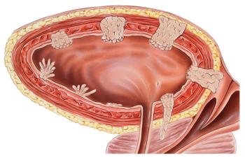
A 67-year-old man, a former smoker, presented with gross hematuria. His initial workup revealed normal renal function, iron deficiency anemia, and increased alkaline phosphatase levels. A CT urogram showed a bladder tumor in the anterior wall and multiple enlarged retroperitoneal lymph nodes. Two vertebral metastases (at T10 and L2) were seen on a bone scan. He underwent a transurethral resection of the bladder, and the pathology report revealed muscle-invasive urothelial carcinoma.
He was treated upfront with cisplatin and gemcitabine. After receiving the first 3 cycles of this regimen, imaging studies demonstrated stable disease. However, upon completion of 6 cycles, he developed mild bone pain; follow-up CT and bone scans revealed an increase in the number and size of the enlarged retroperitoneal lymph nodes, a new right adrenal gland metastasis, and a new bone lesion in the 10th rib compatible with disease progression.
Immunohistochemistry (IHC) of paraffin-embedded bladder tumor tissue revealed > 10% expression of programmed death ligand 1 (PD-L1) in tumor cells and infiltrating immune cells. His performance status was good (Eastern Cooperative Oncology Group [ECOG] performance status score, 1), so he was started on pembrolizumab every 3 weeks as second-line treatment. After 3 cycles, he was found to have a partial response, with his follow-up CT scan showing a 50% decrease in size of the right adrenal gland metastasis and the retroperitoneal lymph node conglomerate (Figure 1). Additionally, the bone scan showed decreased radiotracer uptake in bone lesions (Figure 2). The patient will continue on treatment with pembrolizumab until further assessment.
Which of the following biomarkers indicated that this patient was a good candidate to receive immunotherapy as a second-line therapy after his disease had progressed following platinum therapy-and can be used to make treatment decisions in other patients with metastatic urothelial carcinoma in this setting?
A. PD-L1 expression assessed by IHC on infiltrating immune cells > 5%
B. PD-L1 expression assessed by IHC on tumor cells and infiltrating immune cells > 5%
C. PD-L1 expression assessed by IHC on tumor cells and infiltrating immune cells > 10%
D. There is currently no reliable biomarker
KEY POINTS
- Five immune checkpoint inhibitors have recently been approved for second-line treatment of urothelial carcinoma based on their good response rates and toxicity profiles. Pembrolizumab has demonstrated an overall survival benefit compared with investigator's choice of chemotherapy in a phase III randomized trial.
- Currently, there is no reliable biomarker for selecting candidates for anti–PD-1/PD-L1 agents in this setting. Patients must be considered eligible based on their performance status and prognostic risk factors; PD-1/ PD-L1 testing is not needed.
Correct Answer: D
Discussion
There is currently no reliable biomarker for selecting candidates for programmed death 1 (PD-1)/PD-L1–targeted immunotherapy as second-line therapy in metastatic urothelial carcinoma. Patients are eligible for immune checkpoint inhibitor therapy regardless of their PD-L1 status. The IHC assay, and whether testing should be performed in tumor cells, infiltrating immune cells, or both, have not yet been standardized; the optimal cutoff point for determining PD-L1 status also remains to be defined.
Urothelial carcinoma is the second most common malignancy of the genitourinary system and the sixth most common cancer in the United States. Stage IV metastatic disease is associated with a poor prognosis, with only a 5% survival rate at 5 years.[1,2] Platinum-based chemotherapy leads to high response rates in patients with advanced urothelial carcinoma and has remained the standard of care for first-line systemic treatment, with an expected survival of 9 to 15 months.[3]
Like the patient in this case, most patients will ultimately experience disease progression after platinum-based chemotherapy and will need further therapy.[3] However, there is a lack of effective second-line therapies in this setting. Single-agent chemotherapies used in the second-line setting, such as paclitaxel, docetaxel, gemcitabine, and pemetrexed, have only been tested in phase II studies, in which they have demonstrated low response rates and considerable toxicities.[4] Vinflunine is the only cytotoxic agent to have demonstrated a benefit in a placebo-controlled randomized phase III trial.[5] Nevertheless, the drug showed a disappointing overall response rate (ORR; 8.6%); was not compared against other commonly used chemotherapies, such as taxanes; was never approved in the United States; and in the most recent intention-to-treat analysis, the overall survival (OS) difference (6.9 vs 4.6 months) did not reach statistical significance.[6]
For 30 years, there were no new treatment options approved for urothelial carcinoma. Immune checkpoint inhibitors have recently been found to be very active in neoplasms with high PD-L1 expression and in those with a high burden of somatic mutations.[7] According to the Cancer Genome Atlas Research Network reports, bladder cancer has the third highest rate of mutations, behind lung cancer and melanoma.[8] This fact motivated research studies on checkpoint inhibition after platinum failure, particularly with agents directed against PD-1 or PD-L1. The results of these studies have led to US Food and Drug Administration (FDA) approval of 5 antibodies for this setting within a 1-year timeframe. These include pembrolizumab, nivolumab, atezolizumab, durvalumab, and avelumab.[9-13] The binding of PD-L1 to PD-1 tamps down the immune system. All of the approved drugs prevent the PD-1/PD-L1 interaction and can help restore CD8+ T-cell function targeting tumor cells, thereby facilitating an immunologic anticancer response.[14]
As can be seen in the Table, most of these therapies were evaluated in phase I/II single-arm studies, for which there is a high risk of selection, performance, and detection bias. We decided to prescribe pembrolizumab for our patient, since this drug is the only immunotherapy agent that received regular FDA approval based on mature level 1 evidence demonstrating an OS benefit in metastatic urothelial carcinoma patients after PBCT progression and/or recurrence within 12 months of receiving perioperative chemotherapy for muscle-invasive disease. The study on which the approval was based-KEYNOTE-045-was a phase III, multicenter, open-label randomized trial that compared pembrolizumab vs the investigator’s choice of chemotherapy (single-agent paclitaxel, docetaxel, or vinflunine). The median OS was 10.3 months in the pembrolizumab arm compared with 7.4 months in the chemotherapy arm, with a hazard ratio for death of 0.73 (95% CI, 0.59–0.91; P = .002). The ORR was also higher with pembrolizumab than with cytotoxic treatment (21% vs 11%; P = .0001).[9]
The favorable toxicity profile of pembrolizumab, compared with that of conventional chemotherapy, deserves to be highlighted. Treatment-related adverse events were less common in the PD-1 inhibitor arm than in the chemotherapy arm (60.9% vs 90.2%), and grade 3, 4, or 5 events occurred more frequently in chemotherapy-treated patients (15% vs 49.4%).[9] This is especially important in patients with urothelial carcinoma, given that many of these individuals have comorbid conditions and/or are elderly-and consequently they are often considered unfit for chemotherapy or are unwilling to receive it.[15]
Eligibility for the checkpoint inhibitor trials did not rely on PD-L1 status, but rather on a patient’s performance status and disease burden. In the pembrolizumab trial, patients were eligible for the study if they had an ECOG performance status score of 0, 1, or 2. Patients with a score of 2 were excluded if they had one or more of the established Bellmunt poor prognostic factors (hemoglobin concentration < 10 g/dL, presence of liver metastases, or receipt of the last dose of most recent chemotherapy < 3 months before enrollment). Therefore, the high-risk population was poorly represented in this trial.[9]
In all of the immune checkpoint inhibitor trials, ORRs were reported for the overall population of the study, and also according to PD-L1 status.[7] However, the methods used to evaluate PD-L1 status, the IHC assays utilized, and the cutoff levels varied considerably among studies. In the atezolizumab trial, investigators evaluated PD-L1 expression on tumor-infiltrating immune cells, and the cutoff values were ≥ 1% and ≥ 5%.[11] In the JAVELIN study, which evaluated avelumab, PD-L1 positivity was defined as expression by IHC on ≥ 5% of tumor cells (not immune cells).[13] Durvalumab data defined PD-L1 “high” and “low or absent” expression as ≥ 25% or < 25% of either tumor cells or immune cells staining positive for PD-L1.[12] The CheckMate 275 researchers stratified PD-L1–positive patients into two groups: those with ≥ 1% expression and those with ≥ 5% expression.[10] Finally, the pembrolizumab trial established a PD-L1 combined positive score using a cutoff value of ≥ 10%; they used the percentage of PD-L1–expressing tumor cells and immune cells relative to the total number of cells to calculate the score.[9] As this summary shows, the definition of PD-L1 positivity is very heterogeneous across trials; the standardization of the metrics and procedures used to define the PD-L1 status is an unmet need.
Most trials-but not all-reported better response rates in tumors with high PD-L1 expression. The ORR for atezolizumab-treated patients was 15%; however, response rates were higher in the PD-L1 ≥ 1% and ≥ 5% expression groups-18% (P < .0001) and 27% (P = .0004), respectively.[11] Avelumab achieved a 25% response rate in patients with PD-L1 expression ≥ 5% compared with only 13% in the group with < 5% expression.[13] Durvalumab data showed an 18% ORR; with this agent, responses were seen regardless of PD-L1 expression-27.6% (95% CI, 19.0%–37.5%) and 5.1% (95% CI, 1.4%–12.5%) in patients with high and low or negative expression of PD-L1, respectively.[12] With nivolumab, response rates were 25% in patients with tumor PD-L1 expression ≥ 1% (95% CI, 17.7%–33.6%) and 15.8% in patients with PD-L1 expression < 1% (95% CI, 10.3%–22.7%).[10] The pembrolizumab trial reported similar results in the population of patients who had a PD-L1 combined positive score ≥ 10% and in those with a combined positive score < 10%.[9] Despite a trend toward better outcomes in patients with high PD-L1 expression, the benefit was seen in patients with a wide variety of expression levels.
In the present case, we reported the PD-L1 expression result, since it was done as part of a validation study; however, it was not needed to devise the treatment plan. Currently, PD-L1 expression should not be used as a biomarker for the selection of patients for second-line treatment with PD-1/PD-L1 antibodies.
Having a predictive marker of response for oncologic treatment is desirable, since it helps to optimize decisions in clinical practice. Most efforts in urothelial carcinoma have been focused on PD-L1 expression.[16] However, it is unlikely that evaluation of PD-L1 status as measured by IHC at a single time point would be sufficient to predict response to immunotherapy treatments, since PD-L1 expression on tumor cells and immune cells is a dynamic process. In addition, external factors, such as concomitant therapies (chemotherapy, radiation, or antiangiogenic agents), may induce PD-L1 expression and further affect the response to immunotherapeutic agents.[17]
In conclusion, predictive biomarkers of therapeutic efficacy of PD-1/PD-L1 inhibitors in advanced urothelial carcinoma are lacking. Currently, patients must be considered candidates for immunotherapeutic agents based on their performance status and prognostic risk factors, regardless of their PD-L1 status.
Financial Disclosure:Dr. MarÃa Bourlon has served as a speaker and a member of the advisory board for Bristol-Myers Squibb. The other authors have no significant financial interest in or other relationship with the manufacturer of any product or provider of any service mentioned in this article.
Acknowledgments:The authors would like to thank the Aramont Foundation and Canales de Ayuda A.C. for their support in research activities in Urologic Oncology at Instituto Nacional de Ciencias Médicas y Nutrición Salvador Zubirán.
E. David Crawford, MD, serves as Series Editor for Clinical Quandaries. Dr. Crawford is Professor of Surgery, Urology, and Radiation Oncology, and Head of the Section of Urologic Oncology at the University of Colorado School of Medicine; Chairman of the Prostate Conditions Education Council; and a member of ONCOLOGY's Editorial Board.
If you have a case that you feel has particular educational value, illustrating important points in diagnosis or treatment, you may send the concept to Dr. Crawford at david.crawford@ucdenver.edu for consideration for a future installment of Clinical Quandaries.
References:
1. National Cancer Institute. SEER cancer stat facts: bladder cancer. http://seer.cancer.gov/statfacts/html/urinb.html. Accessed January 10, 2018.
2. American Cancer Society. Cancer facts & figures 2015. https://www.cancer.org/content/dam/cancer-org/research/cancer-facts-and-statistics/annual-cancer-facts-and-figures/2015/cancer-facts-and-figures-2015.pdf. Accessed January 10, 2018.
3. Ortmann CA, Mazhar D. Second-line systemic therapy for metastatic urothelial carcinoma of the bladder. Future Oncol. 2013;9:1637-51.
4. Raggi D, Miceli R, Sonpavde G, et al. Second-line single-agent versus doublet chemotherapy as salvage therapy for metastatic urothelial cancer: a systematic review and meta-analysis. Ann Oncol. 2016;27:49-61.
5. Bellmunt J, Theodore C, Demkov T, et al. Phase III trial of vinflunine plus best supportive care compared with best supportive care alone after a platinum-containing regimen in patients with advanced transitional cell carcinoma of the urothelial tract. J Clin Oncol. 2009;27:
4454-61.
6. Bellmunt J, Fougeray R, Rosenberg JE, et al. Long-term survival results of a randomized phase III trial of vinflunine plus best supportive care versus best supportive care alone in advanced urothelial carcinoma patients after failure of platinum-based chemotherapy. Ann Oncol. 2013;24:1466-72.
7. Gwynn ME, DeRemer DL. The emerging role of PD-1/PD-L1-targeting immunotherapy in the treatment of metastatic urothelial carcinoma. Ann Pharmacother. 2018;52:60-8.
8. Cancer Genome Atlas Research Network. Comprehensive molecular characterization of urothelial bladder carcinoma. Nature. 2014;507: 315-22.
9. Bellmunt J, de Wit R, Vaughn DJ, et al. Pembrolizumab as second-line therapy for advanced urothelial carcinoma. N Engl J Med. 2017;376: 1015-26.
10. Sharma P, Retz M, Siefker-Radtke A, et al. Nivolumab in metastatic urothelial carcinoma after platinum therapy (CheckMate 275): a multicentre, single-arm, phase 2 trial. Lancet Oncol. 2017;18:312-22.
11. Rosenberg JE, Hoffman-Censits J, Powles T, et al. Atezolizumab in patients with locally advanced and metastatic urothelial carcinoma who have progressed following treatment with platinum-based chemotherapy: a single-arm, multicentre, phase 2 trial. Lancet. 2016;387:1909-20.
12. Powles T, O’Donnell PH, Massard C, et al. Efficacy and safety of durvalumab in locally advanced or metastatic urothelial carcinoma: updated results from a phase 1/2 open-label study. JAMA Oncol. 2017;3:e172411.
13. Apolo AB, Infante JR, Balmanoukian A, et al. Avelumab, an anti-programmed death-ligand 1 antibody, in patients with refractory metastatic urothelial carcinoma: results from a multicenter, phase Ib study. J Clin Oncol. 2017;35:2117-24.
14. Maleki Vareki S, Garrigos C, Duran I. Biomarkers of response to PD-1/PD-L1 inhibition. Crit Rev Oncol Hematol. 2017;116:116-24.
15. Chen GJ, Galsky MD, Latini DM, Sonpadve G. Patterns of chemotherapy and survival in elderly patients with advanced bladder cancer: a large Medicare database study. J Clin Oncol. 2013;31(15 suppl):abstr 4551.
16. Katz H, Wassie E, Alsharedi M. Checkpoint inhibitors: the new treatment paradigm for urothelial bladder cancer. Med Oncol. 2017;34:170.
17. Gartrell BA, He T, Sharma J, Sonpavde G. Update of systemic immunotherapy for advanced urothelial carcinoma. Urol Oncol. 2017;35:678-86.


