Oncology NEWS International
- Oncology NEWS International Vol 7 No 8
- Volume 7
- Issue 8
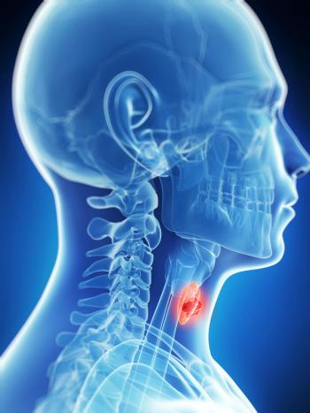
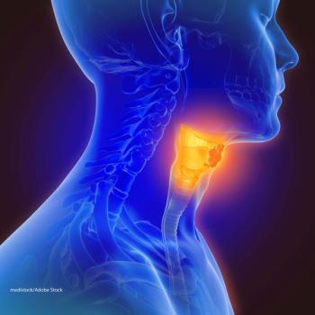
PET for Preoperative Node Staging in Head and Neck Cancer
TORONTO--Therapeutic strategy in patients with head and neck cancer is sometimes based on the staging of regional lymph nodes. However, standard imaging techniques such as computed tomography (CT) and magnetic resonance imaging (MRI) cannot differentiate between inflammation and metastasis in an enlarged lymph node and may not discover distant disease.
TORONTO--Therapeutic strategy in patients with head and neck cancer is sometimes based on the staging of regional lymph nodes. However, standard imaging techniques such as computed tomography (CT) and magnetic resonance imaging (MRI) cannot differentiate between inflammation and metastasis in an enlarged lymph node and may not discover distant disease.
"Correct lymph node staging in squamous cell carcinoma is of most importance in the selection of adequate treatment," Roland Lietzenmayer, MD, said at the Society of Nuclear Medicine meeting. "Currently, lymph node staging of the head and neck area with CT and MRI lacks sensitivity and specificity to tumor infiltration. In addition, tumor cells may also have infiltrated normal-sized lymph nodes, which would not be suspected with conventional imaging techniques."
Dr. Lietzenmayer presented data from a study headed by Dr. Marcus Müller-Berg and performed at Eberhard-Karls-University, Tübingen, Germany.
To evaluate the sensitivity of whole-body fluorine-18-fluorodeoxyglucose positron emission tomography (FDG-PET) in lymph node staging, 29 patients with suspected squamous cell carcinoma were evaluated with FDG-PET, CT, and MRI within 10 to 14 days before therapy.
PET imaging detected all malignant regions via increased focal FDG uptake, for a sensitivity of 100%. Sensitivity with CT was 71% and with MRI, 94%. PET also had higher specificity (97% vs 50% for CT and 58% for MRI). The positive predictive value for PET was 65% vs 27% and 35% for CT and MRI, respectively. Negative predictive value was 100% for PET, 87% for CT, and 98% for MRI.
"Our data indicate that FDG-PET is highly reliable in the preoperative staging of squamous cell carcinoma of the head and neck, and should be performed routinely in these patients," Dr. Lietzenmayer said. He added that if the high negative predictive value seen with PET is confirmed, the technique could potentially be used to individualize surgical treatment.
Also, whole-body FDG-PET imaging might replace other diagnostic procedures, for example, endoscopy to exclude distant metastases or second malignancies. However, he said, CT and MRI scans will still be necessary to provide anatomic information for the surgeons.
Articles in this issue
over 27 years ago
Taxol, Gemzar, Other Therapies Studied in Metastatic Bladder Cancerover 27 years ago
Medicare+Choice Opens Certificationover 27 years ago
Consumer Advocates Win a Voice in NCI Programsover 27 years ago
Paclitaxel Shows Antiangiogenesis Effects in Mouse Modelover 27 years ago
Henney Nominated for FDA Commissionerover 27 years ago
FDA Approves ImageChecker Computer Systemover 27 years ago
Pittsburgh Seeks Breast Cancer Patients for Study on Copingover 27 years ago
Octreotide Imaging Improves Staging of Hodgkin’s Diseaseover 27 years ago
Tobacco Contributions Are Linked to Votes Against Tobacco BillNewsletter
Stay up to date on recent advances in the multidisciplinary approach to cancer.


