- ONCOLOGY Vol 10 No 6
- Volume 10
- Issue 6
Clinical Manifestations of Kaposi's Sarcoma
Major investigative studies have yielded considerable data on various aspects of Kaposi's sarcoma (KS), including cellular and molecular events leading to the development of HIV-associated KS, histopathologic features, clinical
ABSTRACT: Major investigative studies have yieldedconsiderable data on various aspects of Kaposi's sarcoma (KS),including cellular and molecular events leading to the developmentof HIV-associated KS, histopathologic features, clinical characteristics,as well as epidemiologic patterns. Recent developments in researchon KS are summarized, and a coherent model for its pathogenesisis proposed. [ONCOLOGY 10(Suppl):13-23, 1996]
The outbreak of Kaposi's sarcoma (KS) and Pneumocystis cariniipneumonia (PCP) among young, previously healthy, homosexual menin early 1981 marked the initial recognition of the acquired immunedeficiency syndrome (AIDS) [1,2]. KS was among the first AIDS-definingconditions and has remained one of the major cancers associatedwith human immunodeficiency virus-type 1 (HIV-1) infection. Thesudden increase in the incidence of KS has provided considerablenew information. This article summarizes recent developments inresearch on KS and proposes a coherent model for its pathogenesis.
In 1872, Moritz Kaposi first described this disease as "multipleidiopathic pigmented hemangiosarcoma"[3]. He described thecondition as localized, nodular, brown-red to blue-red tumorsthat appear first on the soles of the feet and then on the hands.He was also aware of the multifocal nature of the disease, theoccurrence of visceral involvement, and its vascular nature.
This form of KS, known as classic KS, occurs most commonly inEastern Europe and North America among older men of Italian orJewish ancestry [4]. In the 1950s and 1960s, a more aggressiveform of KS that occurred in younger individuals was describedin central Africa, accounting for 9% of all cancers in Uganda5].In the mid-1970s, KS was reported among a new group of patients,those receiving immunosuppressive therapy for renal transplantation[6,7].
In early 1980, outbreaks of KS were reported among young homosexual/bisexualmen in New York and San Francisco who engaged in promiscuous sexualactivity and had signs of cellular immune defects [1,2,8-11].
Prior to the AIDS epidemic, KS was considered a rare disease [4].The annual incidence of classic KS in the United States was estimatedto be 0.021 to 0.061 per 100,000 people [4,12,13]. Although thisform of KS is seen mostly in persons of Italian or Jewish descent,it has been observed in other groups, including three personsof Eskimoan descent [14] and a large group of people in Swedenand Norway, where there is only a small Jewish population [15,16].A twofold increase in the incidence of classic KS in Sweden duringthe late 1960s was reported in the late 1980s [15]. Other clustersof classic KS have been reported from the Mediterranean islandof Sardinia and on the Peloponnesian peninsula.
The literature indicates a male to female ratio of 15:1 for classicKS, but recent data suggest a ratio of 2:1 to 3:1[4,17]. ClassicKS is most common in black African people [12,13,18,19]. It accountsfor 1.3% of all cancers in Durban, South Africa, 4% in Tanzania,and 9% to 10% in equatorial countries in Africa, such as Ugandaand the Congo [18,19].
Because of the increase in the number of organ transplants andthe more frequent use of immunosuppressive therapy for autoimmunedisorders, iatrogenically induced KS has been reported with increasingfrequency. In organ-transplant recipients, the overall prevalenceof KS is 0.52% (1.24% for liver, 0.45% for kidney, and 0.41% forheart) [20].
It has been established that KS occurs primarily in homosexual/bisexualmen. To a much lesser degree, it also occurs in female partnersof bisexual men and in women from certain regions of the Caribbeanand Africa who acquired HIV infection through heterosexual contact[21]. Other groups at risk of AIDS are reported to develop KS,but to a lesser degree. Approximately 95% of all reported casesof AIDS-associated KS in the United States have been in homosexualand bisexual men, representing 26% of all homosexual men withAIDS. In comparison, AIDS-associated KS is reported in 9% of Haitians,3% of heterosexual intravenous drug abusers, 3% of transfusionrecipients, 3% of women with AIDS, 3% of children with AIDS, and1% of persons with hemophilia. So far, AIDS-associated KS hasnot been seen in women with AIDS who acquired HIV infection throughheterosexual contacts with men with transfusion-acquired AIDSor with iatrogenically infected persons with hemophilia [21].
The incidence of AIDS-associated KS is reported to be quite highamong homosexual/bisexual men who meet their partners in placeswhere anonymous sex is common, such as bath houses. Specifically,sexual practices involving rimming (oral/anal contact) and fisting(insertion of a hand or foot into a partner's rectum) were morecommonly practiced among homosexual/bisexual men who developedAIDS-associated KS.
There has been a steady decline in the incidence of KS since themid-1980s (Table 1). In 1992, it was reported that only 10% ofall patients with AIDS had KS. Since the beginning of the epidemic,the overall incidence of AIDS-associated KS is 13%[22]. Recentchanges in the definition of AIDS-defining conditions will helpreduce the ratio of AIDS-associated KS. This trend has been thesubject of several reviews and discussions [21,23]. One explanationmay be the decreased use of unlabeled amyl nitrite, an inhalantrecreational drug and a possible mitogen [22,24]. Other epidemiologicstudies suggest that changes in certain sexual practices may accountfor the decreasing incidence of KS [25,26].
This steady decline in the incidence of AIDS-associated KS hasbeen paralleled by a decrease in the incidence of seroconversionin homosexual men. Thus, it is believed that changes in the sexualbehavior of homosexual/bisexual men and avoidance of high-riskbehaviors for contracting HIV have been contributing factors.
AIDS-associated KS has been reported in the pediatric population,mostly in the form of lymphadenopathic KS, which is usually detectedat autopsy [27,28]. Mucocutaneous KS has also been reported inchildren who developed HIV infection via blood transfusions [29,30].
In Africa, AIDS-associated KS appears to be transmitted by heterosexualcontact and has an approximately equal incidence among men andwomen [31]. This is in contrast to the male predominance observedin all other populations, and a male-to-female ratio of approximately8:1 for AIDS-associated KS in the United States.
Four different types of KS are recognized: classic, endemic African,iatrogenic, and AIDS-associated. Classic KS usually manifestswith reddish-purple macules, papules, plaques, or nodules. Lesionsmay coalesce to form large plaques and tumors that may producefungating masses with ulceration. Lesions are frequently locatedon the lower extremities but may appear anywhere on the skin ormucous membranes. Older lesions become brown and may develop averrucous and hyperkeratotic surface. The lesions are initiallysoft and spongy but later become hard and solid. Gastrointestinallesions are often asymptomatic in classic KS and are usually detectedonly at autopsy. Nonpitting edema of the lower extremities mayprecede or follow tumor invasion into the superficial and deeplymphatic system. Lymph nodes and internal organs are rarely involved[18]. Classic KS usually runs a benign, protracted course. Rapidprogression with involvement of internal organs is rarely reported.
Clinical forms of African KS have been described as nodular, florid,infiltrative, and lymphadenopathic (Table 2). This classificationis based on the clinicopathologic presentation of the disease.The lymphadenopathic form is usually seen in African childrenand young adults. It manifests mainly with involvement of thelymph nodes and has a rapidly fatal course.
Among recipients of transplants, KS is the second most commontumor after lymphoma. It has been reported to occur more oftenin patients who receive cyclosporine (Sandimmune) as part of theirimmunosuppressive regimen [32]. The mean duration of cyclosporinetherapy before the development of KS is 23 months [32,33]. Iniatrogenic KS, cutaneous involvement is most common; however,lymphatic and visceral dissemination can occur. The disease runsa clinical course similar to that of classic KS, but, in somecases, the course may be more aggressive. Resolution of the diseasehas been reported after withdrawal or reduction of immunosuppressivetherapy [34].
The clinical presentation of AIDS-associated KS is markedly differentfrom that of other forms of the disease. AIDS-associated KS ischaracterized by a multifocal, widespread distribution that mayinvolve any location on the skin or mucous membranes, as wellas the lymph nodes, gastrointestinal tract, and other visceralorgans [35,36]. There is considerable variability in the timingof the initial development of KS in HIV-infected individuals.We have seen patients with AIDS-associated KS who lacked evidenceof immune impairment [35-37]. KS may be the first sign of HIVinfection, especially in populations where HIV testing is notroutinely performed. It can also arise in the later stages ofHIV infection, even up until the last week of life, when patientsare suffering from severe immune deficiency and various opportunisticinfections [37].
Early lesions appear as pink to red macules or tense, purple papules.The lesions appear mostly on the face, especially the nose, eyelids,and ears, and on the trunk. The lower extremities may also beinvolved. The lesions may progress to form plaques, nodules, andtumors, which may eventually erode and ulcerate. The oral mucosamay be involved, with KS lesions appearing on the palate, gingiva,epipharynx, and larynx. Oral cavity involvement is seen in 10%to 15% of patients with AIDS-associated KS. Considerable discomfortand difficulty in eating and swallowing may be caused by oralKS lesions.
Extracutaneous KS is observed at almost every internal site, includingthe gastrointestinal tract, lymph nodes, and lungs. Both the gastrointestinaltract and lymph nodes may be the initial and/or exclusive siteof KS lesions [38-40]. KS lesions are most commonly found in thestomach and duodenum. The lesions are frequently symptomatic,with patients experiencing nausea, ulcerating, bleeding, and ileus.They are localized mostly in the submucosa and are readily visibleby gastroscopy, although they are underdiagnosed by superficialbiopsy.
Patients with pulmonary KS often present with symptoms quite similarto those of PCP, such as intractable cough and respiratory insufficiency[41-43]. Accurate diagnosis is achieved by bronchoscopy, bronchoalveolarlavage, and bronchial biopsy. The prognosis of pulmonary KS ispoor, even with aggressive systemic chemotherapy and radiationtherapy [41-43]. Involvement of bone, subcutaneous tissues andskeletal muscle, bone marrow, peritoneal cavity and omentum, andthe heart is rarely reported [44-49].
The course and progression of KS vary greatly, depending on theclinical form of the disease. The majority of classic KS casesfollow a slow and indolent course. Endemic KS follows a more aggressivecourse, especially in children with the lymphadenopathic form.In AIDS-associated KS, the majority of cases present with a rapidlyprogressive course. The initial localized lesions frequently progressto widespread skin and mucosal involvement and, in some cases,involvement of lymph nodes and solid visceral sites, most commonlythe gastrointestinal tract. Although AIDS-associated KS is moreaggressive than other forms of the disease, patients usually succumbto opportunistic infections. Pulmonary involvement carries a particularlypoor prognosis; although rare, it is a major cause of KS-associatedmortality [50]. At the other extreme are a few cases of indolentdisease that show minimal progression over several years [35,37,51].
Patients with AIDS-associated KS who do not develop opportunisticinfections are estimated to have a survival rate at 28 monthspost-diagnosis of 80%, compared with a survival rate of less than20% in those with opportunistic infections [52]. We have observeda small group of patients with AIDS-associated KS who lived atleast 36 months from the time of biopsy diagnosis of KS withoutdeveloping opportunistic infections. These patients have survivedfrom 142 to 185 months (average, 162 months) (B. S., unpublisheddata, 1995) [35-37]. These long-term survivors of AIDS-associatedKS have normal immune reactivity, thus indicating that immunedeficiency is not a prerequisite for the pathogenesis of KS.
Spontaneous regression has been reported in both classic and AIDS-associatedKS. Regression normally occurs early in the disease and affectsonly some of the lesions. By contrast, in iatrogenic KS, discontinuationof immunosuppressive therapies has been reported to result incomplete regression of the disease [34,53].
Prior to the AIDS epidemic, the most widely used staging systemfor KS was based on the experience from equatorial Africa (Table2) [19]. The initial classification for AIDS-associated KS consideredthe clinical presentation of KS and subtypes A (without systemic"B" symptoms) and B (with systemic "B" symptoms)(!-
A more recent classification system for AIDS-associated KS considersboth clinical and laboratory factors and recognizes the importanceof the T-helper cell count, as did the earlier Walter Reed stagingsystem for HIV disease [55]. A multivariate analysis of 9 variablesperformed on 212 patients showed 3 variables to be prognosticallysignificant: T4 cell counts below 300 cells/mm³, "B"symptoms, and prior or coexisting opportunistic infections.
A staging system based on these three variables, which identifiesfour distinct prognostic groups, has been proposed. The insignificantvariables on multivariate analysis were age, T4/T8 ratio, b2-macroglobulin,acid labile-a interferon (IFN), skin and/or lymph node diseaseonly, and gastrointestinal/palatal lesions. Only on univariateanalysis was the extent of disease, defined as limited skin versusextensive skin and skin and/or lymph node versus gastrointestinaltract and/or palate, found to be significant.
Another proposed staging classification uses three parameters(extent of tumor, immune status, and severity of systemic illness)categorized into good- and poor-risk groups (Table 4) [56]. Onlywith use in prospective trials can the value of predicting treatmentoutcome and survival through these much needed staging systemsbe realized.
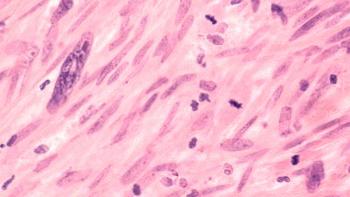
The pathologic process of AIDS-associated KS is reported to startin the mid-dermis and extend upward toward the epidermis. Thecharacteristic histopathologic picture of KS consists of interweavingbands of spindle cells and vascular structures embedded in a networkof reticular and collagen fibers.
Spindle cells may demonstrate a wide range of nuclear pleomorphism.The vascular component may appear as slit-like spaces betweenthe spindle cells or as early, delicate capillaries. Extravasatederythrocytes and hemosiderin-laden macrophages are also present.Mononuclear cell infiltrates are seen, especially in younger lesions.
Based on the quantity of the vascular component, the presenceof nuclear pleomorphism, the number of spindle cells, and theamount of fibrosis in the tumor, three histopathologic patternshave been identified: 1) a mixed cellular pattern containing equalproportions of vascular slits, well-formed vascular channels,and spindle cells; 2) a mononuclear pattern consisting of proliferationof one cell type, usually spindle cells; and 3) an anaplasticpattern characterized by cellular pleomorphism and frequent mitosis.The mixed cellular pattern is seen in all clinical forms of KS,but the anaplastic pattern has been reported only with the floridtype, associated with endemic African KS and some cases of AIDS-KS[19,57].
The histopathologic features of AIDS-associated KS have been thesubject of a large number of reports that include detailed descriptionsof the pathologic changes that occur in various clinical presentations,ie, patch, plaque, and nodular forms of KS [57].
The histopathology of KS in lymph nodes and in viscera is similarto that seen in skin. Foci of KS tumor located in the sinusoidand capsular areas of the lymph nodes and generalized lymphoidhyperplasia are characteristic of KS in the lymph nodes.
In recent years, the increased incidence of KS among HIV-infectedindividuals and advances in biotechnology have made it possibleto investigate the cellular and molecular events leading to thedevelopment and progression of KS. This has resulted in a wealthof knowledge about the pathogenesis of KS.
Cell of Origin--The origin of the cells lining the irregularvascular-like structures and the cells composing the solid tumorfascicle has been the subject of extensive investigations. Histochemicalstaining, cell culture, and ultrastructural studies have all re-sultedin conflicting information (Table 5) [58-70]. Endothelial cells,pericytes, smooth muscle cells, fibroblasts, Schwann cells, andundifferentiated mesenchymal cells have been suspected as KS cells[71-104]; the most recent data support lymphatic endothelial cells.
Long-term culture of AIDS-associated, KS-derived spindle cellshas become possible using conditioned media obtained from CD4+T-cells infected with HTLV-II. [71,77] These long-term culturedcells have been characterized by phenotypic markers and cytochemicalstudies, and it has been shown that the KS spindle cells havefeatures of vascular channel and endothelial cell lineage [67,78,79].The same cells have some features of vascular smooth muscle whengrown on a three-dimensional matrix [69]. A recent study showedthat KS spindle cells share morphologic features with vascularsmooth muscle cells and stain and express the gene for smoothmuscle a-actin [70].
The conclusion drawn from these studies suggested that the spindlecells originate from a mesenchymal precursor cell. In addition,some recent reports suggested involvement of dermal dendrocytes[80-82]. AIDS-associated KS cell lines express several antigenslinking them to dermal dendrocytes, such as factor XIII A, CD4,and CD14 [80-83]. Despite the abundance of new information andthe available technology, the identity of the cells in KS tumorsremains contested.
Although mitotic figures are observed in KS tissues, cytogeneticabnormalities have not been reported, and there is no well-documentedevidence showing KS tumors to metastasize. Some new reports, however,described cultivation of an immortal neoplastic cell line grownfrom an AIDS-KS tissue that had abnormal cytogenetic features.This cell line has been shown to be capable of causing angiogenesis,tumor formation, and metastasis in immunodeficient mice [84,85].Inoculation of this cell line produced tumor at the site of injection,as well as metastatic tumors in the lungs, spleen, pancreas, GItract, and skin. All these tumors have shown a tetraploid karyotypeof human origin [84,85]. These findings suggested that KS cellsmay develop features of malignant neoplastic cells and may becomecapable of metastasising.
Cytokine Cascade in KS--The conditioned media obtainedfrom CD4+ T- cells infected with HTLV-II, which is necessary forthe growth of KS cells, contained basic fibroblast growth factor(FGF), interleukin-1, and a new 30- kD molecule. This new cytokinewas later identified to be a 28- to 36-kD polypeptide producedby activated T- cells and monocytic cells [86]. It was demonstratedthat the new cytokine was actually the same polypeptide previouslydescribed as oncostatin M, a cytokine originally defined by itsability to inhibit the growth of certain melanoma cell lines [86,87].Oncostatin M, which is produced by macrophages and activated T-lymphocytes,has been shown to have a mitogenic effect on KS cells and thusis an autocrine growth factor for KS [87-89]. Oncostatin M actsas a mitogen for cells obtained from KS lesions [88,90] but doesnot induce or maintain the proliferation of normal human endothelialcells [90].
Interleukin-6 (IL-6) has also been reported to function as anautocrine growth factor for AIDS-associated KS [91,92]. It isreported that the a-chain of the IL-6 receptors is expressed inAIDS-associated KS, and an IL-6-dependent autocrine growth loopis present in KS tumors [93]. It has been shown that after exposureto oncostatin M, KS cells acquire the ability to secrete an increasedamount of IL-6 [88,93]. IL-6 is a multifunctional cytokine thathas been shown to be highly inducible at both mRNA and proteinlevels in KS cell cultures by a variety of agents, such as IL-1-b,and tumor necrosis factor-alpha (TNF-a) [92].
Long-term KS cell lines demonstrate that these cells express andproduce a variety of potent biologic activities. KS cell culturesand their products demonstrate growth-promoting activities forKS spindle cells, normal capillary endothelial cells, fibroblasts,and some other mesenchymal cells. They show angiogenic activitiesin CAM assay and nude mice, and they induce a tumor histologicallysimilar to KS, but of mouse origin, when the cells are inoculatedsubcutaneously into nude mice. In addition, they have chemotacticand chemoinvasive activities for KS spindle cells, normal endothelialcells, and fibroblasts [69]; they manifest IL-1 and granulocytemacrophage colony-stimulating factor (GM-CSF)-like activities.77GM-CSF is reported to increase the growth of KS tumors and KScells in cultures [94].
Messenger RNA for platelet-derived growth factor-a (PDGF-a) andIL-6 has also been shown in significant quantities in KS tumors.91In addition, expression of platelet-activating factor, adhesionmolecules, and chemokines by KS cells has been reported [95].
These results have been further confirmed by determining the rateof production and release of these cytokines in conditioned mediaobtained from KS cell cultures using enzyme-linked immuonsorbentassay and radioimmunoprecipitation assays. The sequence analysisof several cDNA clones, Southern blot analysis, and quantitativeslot blot analysis further confirmed high levels of expressionof basic FGF and IL-1-ß by KS cells [96].
It has been demonstrated that KS cells proliferate in responseto the mitogenic effects of recombinant IL-1-a, IL-1-ß,PDGF, and TNF-a, and TNF-ß, whereas smooth muscle cellsrespond to acidic and basic FGF, IL-1, and PDGF [97]. It has beenreported that some of the KS cell cultures express PDGF-b receptors,and these cells are highly responsive to PDGF [98-100]. Theseobservations indicate that KS cells produce cytokines that supporttheir own growth (autocrine) as well as the growth of other cells(paracrine) and that these cytokines may play a major role inthe pathogenesis of AIDS-KS [96,97].
HIV-1 Transactivating (Tat) Gene in KS--The HIV-1 transactivating(Tat) gene and its product, the Tat protein, have beenshown to be important in the pathogenesis of KS [101]. An experimentalmodel has been developed by inserting the Tat gene intothe genome of nude mice. These "transgenic mice" werefound to express Tat-gene mRNA only in skin, and most wenton to develop skin lesions histologically suggestive of earlyKS [102]. The Tat protein induces the expression of inflammatorycytokines [103,104]; supernatant-containing Tat protein stimulatesthe growth of KS cells in culture but not that of normal mesenchymalcells. This growth-promoting effect was inhibited by anti-Tatantibodies and antisense for the Tat protein [101]. Recent studiesdemonstrated the uptake of the Tat protein in tissue culture andthe localization of the Tat protein to the nucleus [105]. Furthermore,it has been shown that basic FGF and the Tat protein have synergisticeffects on KS cells [106]. Thus, the Tat protein may produce theprimary growth stimulus for the development of KS cells in HIV-1-infectedindividuals. The HIV-1 Tat protein has been shown to transactivateTNF-b gene expression through a Tat-like structure [107].
The Tat protein adheres to AIDS-associated KS cells and normalvascular cells through its RGD amino acid sequence. This attachmentis accomplished through a specific interaction with integrin receptorsa5b1 and a5b3. Inflammatory cytokines increase the expressionof both these integrin receptors. Thus, the growth-promoting effectsof the Tat protein on vascular endothelial cells and KS cellsappear to be mediated through the RGD-recognizing integrin sites[108].
Immune Activation--Activation of the immune system is believedto play a role in the pathogenesis of AIDS-associated KS [109-113],as well as classic, endemic, and iatrogenic KS. Patients withclassic KS have high levels of antibodies to cytomegalovirus (CMV)[114,115], which suggests that persistent infection with thisvirus could provide the necessary stimulus for immune activation.Parasitic infection and a variety of other infections could bethe source of continuous antigenic stimulation in endemic AfricanKS. In the case of iatrogenic KS, infections with CMV, Epstein-Barrvirus (EBV), and herpes simplex virus (HSV) may serve as immuneactivators.
In addition, there is immune stimulation from the transplantedtissue alloantigens. Homosexual/bisexual men with AIDS-associatedKS are exposed to chronic antigenic stimuli in the form of multipleviral, bacterial, fungal, and parasitic infections and by spermalloantigens [109,116]. Homosexual men with AIDS-associated KS,compared with a group of healthy homosexual controls, had a muchhigher rate of passive (receptive) anal-genital intercourse associatedwith rectal deposition of the sexual partner's semen, as wellas a higher rate of traumatic sexual practices, such as "fisting,"which would allow entry of organisms and exposure to sperm alloantigens[26]. Furthermore, in the early stages of AIDS-associated KS,paients often do not show immuno suppression, but rather signsof immune activation [35,109,112,113]; patients with AIDS whodevelop KS as their initial manifestation live much longer thanthose presenting with opportunistic infections [52].
Data from several laboratory studies also support the role ofimmune activation in the pathogenesis of KS. Conditioned media,which are necessary for the long-term culture of AIDS-associatedKS cells, have been shown to contain several cytokines normallyproduced by activated immune cells. Conditioned media from phytohemagglutinin-stimulatedperipheral blood mononuclear cells, enriched T cells, and HTLV-I-infectedCD4+ cells have been shown to contain cytokines (IL-1-a, IL-1-ß,IL-6, TNF-a, TNF-ß, GM-CSF, and IL-2) with additive and/orsynergistic mitogenic effects on AIDS-associated KS cells, humanumbilical vein endothelial (hUVE) cells, and adult aortic smoothmuscle cells (aa-SMCs) [117]. It has been demonstrated that hUVEcells and aa-SMCs could become responsive to the mitogenic effectsof the Tat protein (similar to AIDS-associated, KS-derived cells)following exposure to these conditioned media. It has been demonstratedthat TNF-a enhances the progression of KS lesions and augmentsHIV-1 replication in AIDS-associated KS [118-120].
Based on the available data, a hypothetical model for the pathogenesisof AIDS-associated KS has been proposed. In this model, excessiveT-cell activation in HIV-1-infected individuals leads to the releaseof cytokines at levels sufficient to stimulate the proliferationof AIDS-associated KS cells and normal mesenchymal cells; inducesTat protein responsiveness in SMCs and endothelial cells; andactivates HIV-1 replication and Tat gene expression. Inaddition, the extracellular Tat protein, which is released transiently,may stimulate the proliferation of AIDS-associated KS cells andpreactivated mesenchymal cells. It may induce the transactivationof the HIV-1 long terminal repeat and may further amplify HIV-1gene expression and replication. Furthermore, the proliferationof AIDS-associated KS cells leads to a release of cytokines dueto autocrine and paracrine activity, which, in turn, induces neoangiogenesisand the proliferation of mesenchymal cell types [96,97].
In summary, HIV-1 infection and immunostimulation, through theeffects of their extracellular products, might act together toinitiate pathologic molecular and cellular events leading to theproliferation of spindle and mesenchymal cells observed in KSlesions [96,97]. Nevertheless, other factors, such as hormonalinfluences [102], sexual practices [21,26], genetics [121], environmentalfactors [122], and other infectious agents [21], might play rolesin the initiation and development of KS.
The development of KS among some recipients of renal transplantsreceiving immunosuppressive drugs to prevent organ rejection hasgiven rise to the idea that immune deficiency is one of its prerequisites.The appearance of KS as one of the first manifestations of theAIDS epidemic offers further evidence of the possible role ofimmunodeficiency in the development of this disease. However,only a few years into the AIDS epidemic, a small population ofHIV-1-infected patients who developed KS without any clinicopathologicevidence of impaired immunity were identified (B. S., unpublisheddata, 1995). It became apparent that the mechanism by which HIV-1causes KS is different from the mechanism by which it causes immunedeficiency. Furthermore, studies of classic and endemic AfricanKS did not show any true evidence of immune deficiency [123].Based on this information and the recent studies implicating immuneactivation in the development of KS, it no longer appears thatimmune deficiency is necessary for the development of KS. Morelikely, chronic antigenic stimulation and dysregulation of theimmune system are important in the development of AIDS-associatedKS, as well as other forms of this disease.
Available data suggest that the development of KS cannot be simplyexplained by immune stimulation and that a transmissible agentother than HIV may be involved. Many observations have been made:
1) HIV-1 gene products are not found in KS lesions but in theirvicinity;
2) Available clinical and laboratory data indicate that immunedeficiency is not a prerequisite for the development of KS (B.S., unpublished data, 1995) [35,37];
3) Among HIV-infected individuals, the propensity for developingKS does not correlate with the extent of immunosuppression [35,124,125];
4) The observed decline in the number of AIDS-associated KS casesin the United States and Europe is thought to be due to changesin the sexual behavior of HIV-infected homosexual/bisexual men[109,125-130];
5) KS is 300 times more common in patients with AIDS than in iatrogenicallyimmunosuppressed individuals, whereas the incidence of non-Hodgkin'slymphoma in both groups is the same (3%) [30];
6) More than 90% of cases of AIDS-associated KS are seen amonghomosexual/bisexual men [30,31,42,130]; there is a higher incidenceof KS among HIV-negative homosexual men than among other HIV-infectedgroups [131,132];
7) The incidence of AIDS-associated KS is higher among homosexual/bisexualmen who are promiscuous and engage in high-risk sexual activities,such as rimming and fisting, and among receptive sexual partners[31,37,130];
8) Homosexual men who live in New York and San Francisco havea greater risk of developing KS than men who live in the centralstates;
9) AIDS-associated KS is reported to be four times more commonin women with bisexual partners than in women who have had sexwith other HIV-1 seropositive men [30] ;
10) Women from Great Britain who developed AIDS-associated KSare likely to have contracted HIV-1 infection through sexual contactwith bisexual men from the United States [30].
Some data negate the possible involvement of a sexually transmittedagent other than HIV in KS: No single causative agent has beenconsistently isolated in the tissue of KS; it does not explainthe male predominance of African AIDS-associated KS, in whichHIV-1 is transmitted through heterosexual contact; and it doesnot explain the development of classic disease or transplantation-associatedKS, in which there is no evidence of sexual transmission.
Giraldo and co-workers described herpesvirus-like particles intissue cultures derived from patients with classic and endemicAfrican KS [114,115,133-135]. They described a close serologicassociation between CMV and classic KS in North American and Europeanpopulations. Later, CMV-RNA and CMV early antigens were demonstratedin KS tumors and tissue cultures from patients with classic, endemicAfrican, and iatrogenic KS [136-138]. These observations indicatea close association between KS and CMV. CMV may play a part inthe development of KS and may act as a facilitator or promoter,possibly providing the initial cytokine(s) needed for the developmentof a KS lesion. Hepatitis B virus DNA and human papillomavirusDNA and proteins have been observed in patients with AIDS-associatedKS [139-141]. However, neither of these viruses appears to betruly related to KS or its pathogenesis [142].
Retroviruses have also been suspected in AIDS-associated KS, butattempts to isolate a retrovirus from human KS tumors have notbeen successful. Several reports demonstrated retrovirus-likeparticles in KS tumors [143-146]. Considerable excitement accompanieda recent report by Chang et al [147] that described the identificationof herpesvirus-like DNA sequences in AIDS-associated KS. Theseinvestigators employed "representational difference analysis"and were able to isolate unique herpesvirus-like DNA sequencesin more than 90% of KS tumors obtained from AIDS-associated KStissue. These sequences were present in 15% of non-KS tissue DNAsamples from cases with AIDS but were absent in tissue DNA fromnon-AIDS cases. The investigators have reported that these sequencesare homologous to, but distinct from, capsid and tegument proteingenes of the gamma herpesviridae, herpesvirus saimiri, and EBV.
These KS-associated, herpesvirus-like sequences are thought todefine a new human herpesvirus. This initial report was rapidlyfollowed by reports from the same authors as well as other investigatorsindicating the presence of the herpesvirus-like DNA sequencesin other forms of KS (classic, endemic African, and iatrogenicKS) [147-152]. These sequences were found in only 1 of 21 controlskin samples and 3 of 14 uninvolved skin samples from patientswith KS. These DNA sequences were also reported to be presentin AIDS-related, body-cavity-based lymphomas [153]. Body-cavity-basedlymphomas are characterized by pleural, pericardial, or peritoneallymphomatous effusions. More recent reports, however, indicatedthe absence of these fragments in KS cell cultures and their presencein tumors such as squamous cell carcinoma and seborrheic keratosis.Although they appeared at first to be convincing, the presentdata still are not sufficient to prove a causal relationship.Current data are only related to the sequence analysis; the actualvirus has yet to be isolated. The wide distribution of these viralsequences further speaks against their role as an etiologic agent.
The recent interest in KS has resulted in considerable data onvarious aspects of this disease. The epidemiology, pathogenesis,and treatment of KS have been the focus of major investigativework. Many new groups of investigators have become interestedin, and have focused their work on, the study of KS. Undoubtedly,this trend will continue, and many intriguing questions aboutKS will be answered, such as its etiology, its viral status, itscell of origin and possible malignant nature, the role of hormones,and the genetic makeup of the host. A more effective treatmentwill be the ultimate goal. Until then, clinicians must continueto care for their patients and to control their disease. New therapeuticregimens, such as liposomal daunorubicin, have shown considerablepromise toward controlling this disease and reducing its associatedmorbidity.
References:
1. Centers for Disease Control: Pneumocystis pneumonia-Los Angeles.Morbid Mortal Weekly Rep 30:250, 1981.
2. Centers for Disease Control: (Friedman-Kien A, et al): Kaposi'ssarcoma and Pneumocystis pneumonia among homosexual men-New Yorkand California. Morbid Mortal Weekly Rep 30:305-308, 1981.
3. Kaposi M: Classics in oncology: Idiopathic multiple pigmentedsarcoma of the skin. Cancer 31:3-10, 1982.
4. Digiovanna JJ, Safai B: Kaposi's sarcoma: Retrospective studyof 90 cases with particular emphasis on the familial occurrence,ethnic background, and prevalence of other diseases. Am J Med71:779, 1981.
5. Loethe R: Kaposi's sarcoma in Ugandan Africans. Acta PatholMicrobiol Scand 161:1-71, 1963.
6. Myers BD, Kessler E, Levi J, et al: Kaposi's sarcoma in kidneytransplant recipients. Arch Intern Med 133:307, 1974.
7. Shmueli D, Shapira, Yussim A, et al: The incidence of Kaposi'ssarcoma in renal transplant patients and its relation to immunosuppression.Transplant Proc 21:3209, 1989.
8. Haverkos HW, Curran JW: The current outbreak of Kaposi's sarcomaand opportunistic infections. CA Cancer J Clin 32:330, 1982.
9. Gottlieb MS, Schroff R, Schanker HM, et al: Pneumocystit carinipneumonia and mucosal candidiasis in previously healthy men: Evidenceof a new acquired cellular immunodeficiency. N Engl J Med 305:1425,1981.
10. Masur HV, Michelis MA, Greene JB, et al: An outbreak of communityacquired Pneumocystis carinii pneumonia: Initial manifestationof cellular immune dysfunction. N Engl J Med 304:1431, 1981.
11. Siegal FP, Lopez C, Hammer GS, et al: Severe acquired immunodeficiencyin male homosexuals, manifested by chronic perianal ulcerativeherpes simplex lesions. N Engl J Med 305:1439, 1981.
12. Oettle AG: Geographical and racial differences in the frequenciesof Kaposi's sarcoma as evidence of environmental or genetic causes.Acta Un Int Contra Cancrum 18:330-363, 1962.
13. Rothman S: Remarks on sex, age, and racial distribution ofKaposi's sarcoma and on possible pathogenetic factors. Acta UnInt Cancer 18:326, 1962.
14. Mikkelsen F, Nielsen N, Hansen JP: Kaposi's sarcoma in polarEskimos. Acta Derm Venereol 57:539, 1977.
15. Dictor M, Attewell R: Epidemiology of Kaposi's sarcoma inSweden prior to the acquired immunodeficiency syndrome. Int JCancer 42:346, 1988.
16. Klepp O, Dahl O, Stenwig JT, et al: Association of Kaposi'ssarcoma and prior immunosuppressive therapy: A 5 year study ofKaposi's sarcoma in Norway. Cancer 42:2626, 1978.
17. Safai B, Good RA: Kaposi's sarcoma: A review and recent development.CA Cancer J Clin 31:2, 1981.
18. Rothman S: Some clinical aspects of Kaposi's sarcoma in theEuropean and North American populations. Acta Un Int Cancer 18:364,1962.
19. Taylor JF, Templeton AC, Vogel CL, et al: Kaposi's sarcomain Uganda: A clinicopathological study. Int J Cancer 8:122, 1971.
20. Farge D: Kaposi's sarcoma in organ transplant recipients:The Collaborative Transplantation Research Group of Ille de France.Eur J Med 2:339, 1993.
21. Beral V, Peterman TA, Berkelman RL, et al: Kaposi's sarcomaamong persons with AIDS: A sexually transmitted infection? Lancet335:123, 1990.
22. Haverkos HW, Friedman-Kien AE, Drotman DP, et al: The changingincidence of Kaposi's sarcoma among patients with AIDS. J Am AcadDermatol 22:1250, 1990.
23. Elford J, McDonald A, Kaldor J: Kaposi's sarcoma as a sexuallytransmissible infection: An analysis of Australian AIDS surveillancedata. The National HIV Surveillance Committee. AIDS 7:1667, 1993.
24. Archibald CP, Schechter MT, Le TN, et al: Evidence for a sexuallytransmitted cofactor for AIDS-related Kaposi's sarcoma in a cohortof homosexual men (comments). Epidemiology 3:203, 1992.
25. Jaffe HW, Choi K, Thomas PA, et al: National case-controlstudy of Kaposi's sarcoma and Pneumocystis carinii pneumonia inhomosexual men. Part I: Epidemiologic results. Ann Intern Med99:145, 1983.
26. Darrow WW, Jaffe HW, Curran JW: Passive anal intercourse asa risk factor for AIDS in homosexual men. Lancet II:160, 1983.
27. Rogers MF, Thomas PA, Starcher ET, et al: Acquired immunodeficiencysyndrome in children: Report of the Centers for Disease Control.National Surveillance, 1982-1985. Pediatrics 79:1008, 1987.
28. Buch BE, Scott GB, Valdes-Dapena M, et al: Kaposi's sarcomain two infants with acquired immune deficiency syndrome. J Pediatr103:911, 1983.
29. Connor E, Boccon-Gibod L, Joshi V, et al: Cutaneous acquiredimmunodeficiency syndrome-associated Kaposi's sarcoma in pediatricpatients. Arch Dermatol 126:791, 1990.
30. Gutierrex-Ortega P, Hierro-Orozco S, Sanchez-Cisneros R, etal: Kaposi's sarcoma in a 6-day-old infant with human immunodeficiencyvirus. Arch Dermatol 125:432, 1989.
31. Bayley AC, Downing RG, Cheingsong-Popov R, et al: HTLV-IIIserology distinguishes atypical and endemic Kaposi's sarcoma inAfrica. Lancet I:359-361, 1985.
32. Civati G, Busnach G, Brando B, et al: Occurrence of Kaposi'ssarcoma in renal transplant recipients treated with cyclosporine.Transplant Proc 20 (suppl 3):924, 1988.
33. Harwood AR, Osoba SD, Hofstadler SL, et al: Kaposi's sarcomain recipients of renal transplants. Am J Med 67:759, 1979.
34. Zisbrod Z, Haimov M, Schanzer H, et al: Kaposi's sarcoma afterkidney transplantation: Report of complete remission of cutaneousand visceral involvement. Transplantation 30:383, 1980.
35. Safai B, Johnson KG, Myskowski PL, et al: The natural historyof Kaposi's sarcoma in the acquired immunodeficiency syndrome.Ann Intern Med 103:744, 1985.
36. Niedt GW, Schinella RA: Acquired immunodeficiency syndrome:A clinicopathologic study of 56 autopsies. Arch Pathol Lab Med109:727, 1985.
37. Safai B, Sarngadharan MG, Koziner B, et al: Spectrum of Kaposi'ssarcoma in the epidemic of AIDS. Cancer Res 45:4646S-4648S, 1985.
38. Rose HS, Balthazar EJ, Megibow AJ, et al: Alimentary tractinvolvement in Kaposi's sarcoma: Radiographic and endoscopic findingsin 25 homosexual men. Am J Radiol 139:661, 1982.
39. Frager DH, Frager JD, Brandt LJ, et al: Gastrointestinal complicationsof AIDS: Radiologic features. Radiology 158:597, 1986.
40. Galtz RK: Kaposi's sarcoma: Gastrointestinal involvement correlationwith skin findings and immunologic functions. Dig Dis Sci 29:817,1984.
41. Kaplan LD, Hopewell PC, Jaffe H, et al: Kaposi's sarcoma involvingthe lung in patients with the acquired immunodeficiency syndrome.J Acquir Immune Defic Syndr 1:23, 1988.
42. Ognibene FP, Steis RG, Macher AM, et al: Kaposi's sarcomacausing pulmonary infiltrates and respiratory failure in the acquiredimmunodeficiency syndrome. Ann Intern Med 102:471, 1985.
43. Pitchenik AE, Fischl MA, Saldana MJ, et al: Kaposi's sarcomaof the tracheobronchial tree: Clinical, bronchoscopic, and pathologicfeatures. Chest 87:122, 1985.
44. Nicholas CM, Flaitz CM, Hicks MJ: Primary intraosseous Kaposi'ssarcoma of the maxilla in human immunodeficiency virus infection:Review of literature and report of case. J Oral Maxillofac Surg53:325, 1995.
45. Leal R, Lewin M, Ahman I, at el: Peritoneal Kaposi's sarcoma:a cause of ascites in acquired immunodeficiency syndrome. DigDis Sci 39:206, 1994.
46. Levin M, Hertzberg L: Kaposi's sarcoma of the bone marrowpresenting with fever of unknown origin. Med Pediatr Oncol 22:410,1994.
47. Lopez Rubio F, Anguita M, Arizon JM, et al: Visceral Kaposi'ssarcoma mucocutaneous involvement in a heart transplant recipient.J Heart Lung Transplant 13:913, 1994.
48. Lin O, Scholes JV, Lustbader IJ: Chylous ascites resultingfrom Kaposi's sarcoma in an AIDS patient. Am J Gastroenterol 89:2252,1994.
49. Martinez Sapina MJ, Mosquera J, Castro JM, et al: Kaposi'ssarcoma involving bone in an HIV negative patient. Eur J Radiol15:200, 1992.
50. Meduri GU, Stover DE, Lee M, et al: Pulmonary Kaposi's sarcomain the acquired immunodeficiency syndrome. Am J Med 81:11, 1986.
51. Hardy AM: Characterization of long term survivors of acquiredimmunodeficiency syndrome. J AIDS 4:386-391, 1991.
52. Rothenberg R, Woelfel M, Stoneburner R, et al: Survival withthe acquired immunodeficiency syndrome: Experience with 5,833cases in New York City. N Engl J Med 317:1297, 1987.
53. Pilgrim M: Spontaneous manifestation of regression of a Kaposi'ssarcoma under cyclosporin A. Hautarzt 39:368, 1988.
54. Krigel RL, Laubenstein LJ, Muggia FM: Kaposi's sarcoma: Anew staging classification. Cancer Treat Rep 67:531, 1983.
55. Redfield RR, Wright DC, Tramont EC: The Walter Reed stagingclassification for HTLV-III/LAV infection. N Engl J Med 314:131,1986.
56. Krown SE, Metroka C, Wernz JC, et al: Kaposi's sarcoma inthe acquired immune deficiency syndrome: A proposal for uniformevaluation, response, and staging criteria. J Clin Oncol 7:1201,1988.
57. Gottleib GJ, Ackerman AB, eds: Kaposi's Sarcoma: A Text andAtlas. Baltimore, Lea & Febiger, 1988.
58. Pepler WJ: The origin of Kaposi's haemangiosarcoma: A histochemicalstudy. J Pathol Bact 78:553, 1959.
59. Pepler WJ, Theron JJ: An electron microscopic study of Kaposi'shaemangiosarcoma. J Pathol Bact 83:521, 1962.
60. Hashimoto K, Lever WF: Kaposi's sarcoma: Histochemical andelectron microscopic studies. J Invest Dermatol 43:539, 1964.
61. Dayan AD, Lewis PD: Origin of Kaposi's sarcoma from the reticuloendothelialsystem. Nature 2:889, 1967.
62. Harrison AC, Kahn LB: Myogenic cells in Kaposi's sarcoma:An ultrastructural study. J Pathol 124:157, 1978.
63. Nadji M, Morales AR, Ziegles-Weissman J, et al: Kaposi's sarcoma:Immunohistologic evidence for an endothelial origin. Arch PatholLab Med 105:274, 1981.
64. Beckstead JH, Wood GS, Fletcher V: Evidence for the originof Kaposi's sarcoma from lymphatic endothelium. Am J Pathol 119:294,1985.
65. Rutgers JL, Wieczorek R, Bonetti F, et al: The expressionof endothelial cell surface antigens by AIDS-associated Kaposi'ssarcoma: Evidence for a vascular endothelial cell origin. Am JPathol 122:493, 1986.
66. Jones RR, Spaull J, Spry C, et al: Histogenesis of Kaposi'ssarcoma in patients with and without acquired immunodeficiencysyndrome (AIDS). J Clin Pathol 39:742, 1986.
67. Salahuddin SZ, Nakamura S, Biberfeld, et al: Angiogenic propertiesof Kaposi's sarcoma-derived cells after long-term culture in vitro.Science 242:430, 1988.
68. Nickoloff BJ, Griffiths CEM: The spindle-shaped cells in cutaneousKaposi's sarcoma. Am J Pathol 135:793, 1989.
69. Thompson EW, Nakamura S, Shima TB, et al: Supernatants ofacquired immunodeficiency syndrome-related Kaposi's sarcoma cellsinduce endothelial cell chemotaxis and invasiveness. Cancer Res51:2670, 1991.
70. Weich HA, Salahuddin SZ, Nakamura S, et al: AIDS-Kaposi'sderived cells in long-term culture express and synthesize smoothmuscle a-actin. Am J Pathol 139:1251, 1991.
71. Nakamura S, Salahuddin SZ, Biberfeld P, et al: Kaposi's sarcomacells: Long-term culture with growth factor from retrovirus-infectedCD4+ T cells. Science 242:426, 1988.
72. Harrison AC, Kahn LB: Myogenic cells in Kaposi's sarcoma:An ultrastructural study. J Pathol 124:256, 1978.
73. Sterry W, Steigleer G: Kaposi's sarcoma: Venous capillaryhemangioblastoma: A histochemical and ultrastructural study. ArchDermatol Res 266:253, 1979.
74. Witte MH, Witte CL: AIDS-Kaposi's sarcoma complex: Evolutionof a full blown lymphologic syndrome. Lymphology 21:4, 1988.
75. Dorfman RF: Kaposi's sarcoma: Evidence supporting its originfrom the lymphatic system. Lymphology 21:45, 1988.
76. Dictor M: Vascular remodeling in Kaposi's sarcoma and avianhemangiomatosis: Relation to the vertebrate lymphatic system.Lymphology 21:53, 1988.
77. Ensoli B, Nakamura S, Salahuddin SZ, et al: AIDS-Kaposi'ssarcoma-derived cells express cytokines with autocrine and paracrinegrowth effects. Science 243:223, 1989.
78. Benelli R, Repetto L, Carlone S, et al: Establishment andcharacterization of two new Kaposi's sarcoma cell cultures froman AIDS and a non-AIDS patient. Res Virol 145:252, 1994.
79. Herndier BG, Werner A, Arnstein P, et al: Characterizationof a human Kaposi's sarcoma cell line that induces angiogenictumors in animals. AIDS 8:575, 1994.
80. Huang YQ, Friedman Kien AE, Li JJ, et al: Cultured Kaposi'ssarcoma cell lines express factor X111a, CD14, and VCAM-1, butnot factor VIII or ELAM-1 (published erratum. Arch Dermatol 12:1622,1993.
81. Kanitakis J, Roca Miralles M: Factor-X111a-expressing dermaldendrocytes in Kaposi's sarcoma: A comparison between classicaland immunosuppression-associated types. Virchows Arch A PatholAnat Histopathol 420:227, 1992.
82. Kumar D, Sanchez RL, Kumar S: Dendrocyte population in cutaneousand extracutaneous Kaposi's sarcoma. Am J Dermatopathol 14:298,1992.
83. Regezi JA, MacPhail LA, Daniels TE, et al: Human immunodeficiencyvirus-associated oral Kaposi's sarcoma: A heterogeneous cell populationdominated by spindle-shaped endothelial cells. Am J Pathol 143:240,1993.
84. Lunardi-Iskander Y, Gill P, Lam VH, et al: Isolation and characterizationof an immortal neoplastic cell line (KSY-1) from AIDS-associatedKaposi's sarcoma. J Natl Cancer Inst 87:974, 1995.
85. Lunardi-Iskander Y, Bryant JL, Zeman RA, et al: Tumorigenesisand metastasis of neoplastic Kaposi's sarcoma cell line in immunodeficientmice blocked by a human pregnancy hormone. Nature 375:64, 1995.
86. Nair BC, DeVico AL, Nakamura S, et al: Identification of amajor growth factor for AIDS-Kaposi's sarcoma cells as oncostatinM. Science 255:1430, 1992.
87. Zarling JM, Shoyab M, Marquardt H, et al: Oncostatin M: Agrowth regulator produced by differentiated histiocytic lymphomacells. Proc Natl Acad Sci USA 83:9739, 1986.
88. Miles SA, Martinez-Maza O, Rezai A, et al: Oncostatin M asa potent mitogen for AIDS-Kaposi's sarcoma-derived cells. Science255:1432, 1992.
89. Cai J, Gill PS, Masood R, et al: Oncostatin M is an autocrinegrowth factor in Kaposi's sarcoma. Am J Pathol 145:74, 1994.
90. Brown TJ, et al: Regulation of IL-6 expression by OncostatinM. J Immunol 147:2175, 1991.
91. Miles SA, Rezai AR, Salazar-Gonzalez JF, et al: AIDS Kaposi'ssarcoma-derived cells produce and respond to interleukin-6. ProcNatl Acad Sci USA 87:4068, 1990.
92. Yang J, Hagan MK, Offermann MK: Induction of IL-6 gene expressionin Kaposi's sarcoma cells. J Immunol 152:943, 1994.
93. Miles SA: Pathogenesis of human immunodeficiency virus-relatedKaposi's sarcoma. Curr Opin Oncol 4:875, 1992.
94. Hermans P, Gori A, Lemone M, et al: Possible role of granulocytemacrophage colony stimulating factor (GM-CSF) on the rapid progressionof AIDS-related Kaposi's sarcoma lesions in vivo. Br J Haematol87:413, 1994.
95. Sciacca FL, Sturzl M, Bussolino F, et al: Expression of adhesionmolecules, platelet-activating factor, and chemokines by Kaposi'ssarcoma cells. J Immunol 153:4816, 1994.
96. Ensoli B, Salahuddin SZ, Gallo RC: AIDS-associated Kaposi'ssarcoma: A molecular model for its pathogenesis. Cancer Cells1:93, 1989.
97. Ensoli B, Barillari G, Gallo RC: Pathogenesis of AIDS-associatedKaposi's sarcoma. Hematol Oncol Clin North Am 5(2):281, 1991.
98. Roth WK, Werner S, Schirren CG, et al: Depletion of PDGF fromserum inhibits growth of AIDS-related and sporadic Kaposi's sarcomacells in culture. Oncogene 4:483, 1989.
99. Sturzl M, Roth WK, Brockmeyer NH, et al: Expression of platelet-derivedgrowth factor and its receptor in AIDS-related Kaposi sarcomain vivo suggests paracrine and autocrine mechanisms of tumor maintenance.Proc Natl Acad Sci USA 89:7046, 1992.
100. Pistritto G, Ventura L, Mores N, et al: Regulation of PDGF-Bin PDGF receptor expression in the pathogenesis of Kaposi's sarcomain AIDS. Antibiot Chemother 46:73, 1994.
101. Ensoli B, Gendelman R, Markham P, et al: Tat protein of HIV-1stimulates growth of cells derived from Kaposi's sarcoma lesionsof AIDS patients. Nature 345:84, 1990.
102. Vogel J, Hinrichs SH, Reynolds RK, et al: The HIV tat geneinduces dermal lesions resembling Kaposi's sarcoma in transgenicmice. Nature 335:606, 1988.
103. Buonaguro L, Barillari G, Chang HL, et al: Effects of thehuman immunodeficiency virus type 1 Tat protein on the expressionof inflammatory cytokines. J Virol 66:7159, 1992.
104. Ensoli B, Buonaguro L, Barillari G, et al: Release, uptake,and effects of extracellular HIV-1 Tat protein on cell growthand viral transactivation. J Virol 67: 277-287, 1993.
105. Frankel AD, Paba CO: Cellular uptake of the Tat protein fromhuman immunodeficiency virus. Cell 55:1189, 1988.
106. Ensoli B, Gendelman R, Markham P, et al: Synergy betweenbasic fibroblast growth factor and HIV-1 Tat protein in inductionof Kaposi's sarcoma. Nature 371:674, 1994.
107. Buonaguro, L, Buonaguro FM, Giraldo G, et al: The human immunodeficiencyvirus type 1 Tat protein transactivates tumor necrosis factorbeta gene expression through a Tar-like structure. J Virol 68:2677,1994.
108. Barillari G, Gendelman R, Gallo RC, et al: The Tat proteinof human immunodeficiency virus type 1, a growth factor for AIDSKaposi sarcoma and cytokine-activated vascular cells, inducesadhesion of the same cell types by using integrin receptors recognizingthe RGD amino acid sequence. Proc Natl Acad Sci USA 90:7941, 1993.
109. Jacobson LP, Munos A, Fox R, et al: Incidence of Kaposi'ssarcoma in a cohort of homosexual men with human immunodeficiencyvirus type 1. J AIDS 3(suppl 1):S24-S31, 1990.
110. Rabkin CS, Goedert JJ, Biggar RJ, et al: Kaposi's sarcomain three HIV-1-infected cohorts. J AIDS 3(suppl 1):S38-S43, 1990.
111. Mitsuyasu RT, Taylor JMG, Glaspy J, et al: Heterogeneityof epidemic Kaposi's sarcoma. Cancer 57:1657-1661, 1986.
112. Ballard HS: Dissemination of Kaposi's sarcoma without lymphocyteabnormalities. Arch Intern Med 145:547, 1985.
113. Lane HC, Masur H, Gelman EP, et al: Correlation between immunologicfunction and clinical subpopulations of patients with the acquiredimmunodeficiency syndrome. Am J Med 78:417, 1985.
114. Giraldo G, Beth E, Kourilsky FM, et al: Antibody patternsto herpesvirus in Kaposi's sarcoma. I: Serological associationof European Kaposi's sarcoma with cytomegalovirus. Int J Cancer15:839, 1975.
115. Giraldo G, Beth E, Henle W, et al: Antibody patterns to herpesvirusin Kaposi's sarcoma. II: Serological association of American Kaposi'ssarcoma with cytomegalovirus. Int J Cancer 22:126, 1978.
116. Mavligit GM, Talpaz M, Hsia FT, et al: Chronic immune stimulationby sperm alloantigens. JAMA 251:237, 1984.
117. Barillari G, Buonaguro L, Fiorelli V, et al: Effects of cytokinesfrom activated immune cells on vascular cell growth and HIV-1gene expression: Implications for AIDS-Kaposi's sarcoma pathogenesis.J Immunol 149:3727, 1992.
118. Duh EJ, Maury WJ, Folks TM, et al: Tumor necrosis factora activates human immunodeficiency virus type 1 through inductionof nuclear factor binding to the NF-kB sites in the long terminalrepeat. Microbiology 86:5974, 1989.
119. Poli G, Kinter A, Justement JS, et al: Tumor necrosis factora functions in an autocrine manner in the induction of human immunodeficiencyvirus expression. Immunology 87:782, 1990.
120. Osborn L, Kunkel S, Nabel GJ: Tumor necrosis factor a andinterleukin-1 stimulate the human immunodeficiency virus enhancerby activation of the nuclear factor kB. Proc Natl Acad Sci USA86:2336-2340, 1989.
121. Pollack MS, Safai B, DuPont B: HLA-DR5 and DR2 are susceptibilityfactors for acquired immunodeficiency syndrome with Kaposi's sarcomain different ethnic subpopulations. Dis Markers 1:135-139, 1983.
122. Haverkos HW, Pinsky PF, Drotman DP, et al: Disease manifestationamong homosexuals with acquired immunodeficiency syndrome: A possiblerole of nitrites in Kaposi's sarcoma. Sex Transm Dis 12:203, 1985.
123. Safai B, Mike V, Giraldo G, et al: Association of Kaposi'ssarcoma with second primary malignancies. Cancer 45:1472, 1980.
124. Goedert JJ, Biggar RJ, Melbye M, et al: Effect of T4 countand cofactors on the incidence of AIDS in homosexual men infectedwith human immunodeficiency virus. JAMA 257:331, 1987.
125. Beral V, et al: Risk of Kaposi's sarcoma in AIDS patientsin the British Isles: Is it increased if sexual partners comefrom the USA or Africa? Br Med J 325:624, 1991.
126. Piot P, Quinn TC, Taelman H, et al: Acquired immunodeficiencysyndrome in a heterosexual population in Zaire. Lancet 2:65, 1984.
127. Drew WL, Mills J, Hauer LB, et al: Declining prevalence ofKaposi's sarcoma in homosexual AIDS patients paralleled by fallin cytomegalovirus transmission (letter). Lancet 327:66, 1988.
128. Detels R, English P, Visscher BR, et al: Seroconversion,sexual activity, and condom use among 2915 HIV-seronegative menfollowed for up to 2 years. J Acquir Immune Defic Syndr 2:77,1989.
129. Lifson AR, Darrow WW, Hessol NA, et al: Kaposi's sarcomaamong homosexual and bisexual men enrolled in the San FranciscoCity Clinic Cohort Study. J Acquir Immune Defic Syndr 3(suppl1):S32, 1990.
130. Beral V, et al: Risk of Kaposi's sarcoma and sexual practicesassociated with faecal contact in homosexual or bisexual men withAIDS. Lancet 339:632, 1992.
131. Safai B, Peralta H, Menzies K, et al: Kaposi's sarcoma amongHIV-seronegative high risk population. VII International Conferenceon AIDS. Florence, Italy, June 1991.
132. Friedman-Kien AE, Saltzman BR, Cao Y, et al: Kaposi's sarcomain HIV-negative homosexual men. Lancet 335:168, 1990.
133. Giraldo G, et al: Kaposi's sarcoma: A new model in the searchfor viruses associated with human malignancies. J Natl CancerInst 49:1495, 1972.
134. Giraldo G, et al: Herpes-type virus particles in tissue cultureof Kaposi's sarcoma from different geographic regions. J NatlCancer Inst 49:1509, 1972.
135. Giraldo G, Beth E, Huang ES: Kaposi's sarcoma and its relationshipto cytomegalo-
virus (CMV). III: CMV DNA and CMV early antigen in Kaposi's sarcoma.Int J Cancer 26:23, 1980.
136. Boldogh I, Beth E, Huang ES, et al: Kaposi's sarcoma. IV:Detection of CMV-DNA, CMV-RNA, and CMNA in tumor biopsies. IntJ Cancer 28:469, 1981.
137. McDougall JK, Nelson JA, Myerson D, et al: HSV, CMV, andHPV in human neoplasia. J Invest Dermatol 83:72S, 1984.
138. Siegal B, et al: Kaposi's sarcoma in immunosuppression: Possiblythe result of a dual viral infection. Cancer 65:492, 1990.
139. Siddiqui A: Hepatitis B virus DNA in Kaposi's sarcoma. ProcNatl Acad Sci USA 80:4861, 1983.
140. Nickoloff BJ, Huang YQ, Li JJ, et al: Immunohistochemicaldetection of papillomavirus antigens in Kaposi's sarcoma. Lancet339:548, 1992.
141. Huang YQ, Li JJ, Rush MG: HPV-16-related DNA sequences inKaposi's sarcoma.
Lancet 339: 515, 1992.
142. Biggar RJ, Dunsmore N, Kurman RJ, et al: Failure to detecthuman papillomavirus in Kaposi's sarcoma (letter; comment). Lancet339:1604, 1992.
143. Rappersberger K et al: Demonstration of (im)mature oncovirinaein tumour cells from patients with Mediterranean and classic Kaposi'ssarcoma. Arch Dermatol Res 283:58, 1991.
144. Gyorkey F, et al: Retroviruses in Kaposi's sarcoma cellsin AIDS. N Engl J Med 311:1183, 1984.
145. Schenk P, Konrad K: Ultrastructure of Kaposi's sarcoma inacquired immune deficiency syndrome (AIDS). Arch Otorhinolaryngol242:305, 1985.
146. Schenk P: Retroviruses in Kaposi's sarcoma in acquired immunedeficiency syndrome (AIDS). Acta Otolaryngol Stockh 202:295, 1986.
147. Chang Y, Cesarman E, Pessin MS, et al: Identification ofherpesvirus-like DNA sequence in AIDS-associated Kaposi's sarcoma.Science 266:1865, 1994.
148. Huang YQ, Li JJ, Kaplan MH, et al: Human herpesvirus-likenucleic acid in various forms of Kaposi's sarcoma. Lancet 345:759,1995.
149. Jones JL, Hanson DL, Chu SY: AIDS-associated Kaposi's sarcoma(letter). Science 267:1078, 1995.
150. Ambrogiak JA, Blackbourne DJ, Herndier BQ, et al: Herpes-likesequences in HIV-infected and uninfected Kaposi's sarcoma patients.Science 268:582, 1995.
151. Moore PS, Chang Y: Detection of herpesvirus-like DNA sequencesin Kaposi's sarcoma in patients with and those without HIV infection.N Engl J Med 332:1181, 1995.
152. Su I-J, Hsu Y-S, Chang Y-C, et al: Herpesvirus-like DNA sequencein Kaposi's sarcoma from AIDS and non-AIDS patients in Taiwan.Lancet 345:722, 1995.
153. Cesarman E, Chang Y, Moore PS, et al: Kaposi's sarcoma-associatedherpesvirus like DNA sequences in AIDS-related body cavity-basedlymphomas. N Engl J Med 332:1186, 1995.
Articles in this issue
over 29 years ago
AIDS-related Kaposi's Sarcoma: Options for Today and Tomorrowover 29 years ago
New Developments: A Look to the Futureover 29 years ago
The Epidemiologic Scope of Kaposi's Sarcomaover 29 years ago
Current Therapeutic Options for Kaposi's Sarcomaover 29 years ago
Discussionover 29 years ago
Comments on Bone Marrow Transplantation for Multiple Myelomaover 29 years ago
Medicine as Business May Mean More Ethical Challenges for PhysiciansNewsletter
Stay up to date on recent advances in the multidisciplinary approach to cancer.


