- ONCOLOGY Vol 10 No 6
- Volume 10
- Issue 6
New Developments: A Look to the Future
Inflammatory cytokines plus the human immunodeficiency virus Tat protein apparently trigger the development of early Kaposi's sarcoma. Activated spindle cells provide a self-perpetuating, autocrine-supported mechanism for further development of hyperplastic lesions. In more advanced stages, a true neoplastic process may develop. [ONCOLOGY 10(Suppl):34-36, 1996]
ABSTRACT: Inflammatory cytokines plus the human immunodeficiency virus Tat protein apparently trigger the development of early Kaposi's sarcoma. Activated spindle cells provide a self-perpetuating, autocrine-supported mechanism for further development of hyperplastic lesions. In more advanced stages, a true neoplastic process may develop. [ONCOLOGY 10(Suppl):34-36, 1996]
Kaposi's sarcoma (KS) is of special interest not only becauseof its association with the epidemic of human immunodeficiencyvirus (HIV) infection (AIDS-KS), but also because studies of itspathogenesis appear likely to contribute to a deeper understandingof tumor biology, particularly angiogenesis. This is true despitea major paradox: researchers are still uncertain as to whetheror not the four types of KS actually represent a single diseaseand even whether or not KS, which can certainly produce tumors,is a true neoplasm.
KS tumors are characterized by a significant complexity of celltypes. Many normal cells infiltrate into the mass generally referredto as the KS lesion [1-3]. Furthermore, in most patients withthis disorder, it remains difficult to prove that there are trulyclonal, proliferating, malignant cells at any time.
Our group and another group from Israel have established KS celllines that are truly neoplastic; our tumor cell line (KS Y-1,derived from AIDS-KS) [4] produces metastatic disease in mice,whereas the other cell line (derived from a patient with classicKS) is tumorigenic but not metastatic [5]. However, these linesare derived from cells that appear to be quite rare in KS lesions,because many attempts have been made to establish cell lines fromother cell types, with no success. Another possibility, whichmost likely accounts for the majority of KS lesions, is that thesignificant event may be hyperplasia-induced and maintained bythe spindle cell, which secretes or releases cytokines that permitinfiltration and proliferation of other cell types [6-10]. Ourwork has focused on characterizing cells from KS lesions in termsof the phenotype and cytokines they secrete and their abilityto create model KS lesions when inoculated into nude mice. Wehave demonstrated that most of the KS spindle cells that clearlywere not neoplastic [11,12] have the same phenotype as that ofmost of the in situ spindle cells [2,13] and can produce a lesionin mice that is histopathologically indistinguishable from earlyKS [11,12].
The multifocal nature of this tumor raises the question of whetherthis could be hyperplasia, rather than neoplasia, at least inits early stage. If so, the abnormal milieu of HIV-infected individualsmay be permitting an increased local proliferation of a varietyof cell types, depending upon the local environment. If this isthe case, the spindle cell may have become seeded in the bloodand disseminated to sites where it can reside and induce localinflammation-like changes. Neoplastic conversion may be a chancelate-stage phenomenon.
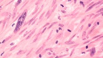
Culture of cells from primary biopsy specimens of patients withKS is achieved by using conditioned media from activated T-cells[11,14,15]. These cells have a spindle-shaped morphology and anactivated endothelial-cell phenotype that is identical to thatof most in situ spindle cells [2,13].
Conversely, normal endothelial cells grown in a medium that containsinflammatory cytokines from activated T-cells give rise to a similaror identical type of spindle-shaped cell [13,16]. In both cases,these cells have receptors for a great number and variety of cytokines.The predominant cytokines involved in producing this activatedcell are interleukin-1 (IL-1), tumor necrosis factor, and gammainterferon. Of importance, these cytokines are the same inflammatorycytokines increased in KS lesions (B.E., unpublished data, 1995).
Once activated in this way, the spindle cell then proliferatesand produces its own cytokines in very large amounts. This isparticularly true of basic fibroblast growth factor (bFGF), whichis not released in significant amounts by normal cells [6]. TheKS spindle cells have receptors for bFGF and produce and releasemore bFGF than any other known cell both in vitro and in vivo[6,17,18]. Basic fibroblast growth factor is also highly expressedby in situ spindle cells of both classic KS and AIDS-KS lesions,inducing autocrinic KS cell growth. Upon release, bFGF promotesangiogenesis and KS-like lesions that are indistinguishable fromthose induced by KS spindle cells or lesions induced by cytokine-activatedendothelial cells [6,17-19].
The spindle cell also produces vascular endothelial cell growthfactor, platelet-derived growth factor, IL-6, granulocyte-macrophagecolony stimulating factor (GM-CSF), and IL-8 [6,8,9,20]. Someof these cytokines also serve an autocrinic function in keepingthe spindle cell functioning. The most important factor, however,appears to be bFGF, because most spindle cell growth can be blockedby polyclonal antibodies to bFGF or antisense oligodeoxynucleotidesdirected against bFGF [6,17].The derivation of the KS spindlecell has been recently elucidated. Most of the current findingsindicate a microvascular endothelial origin in an activated state[2,3,13]. Cultured spindle cells do lack the vascular endothelialcell marker FVIII-RA; however, this molecule is released in thepresence of inflammatory cytokines. In fact, when KS cells arecultured in their absence, the expression of this molecule isregained [13]. In addition, these cells present other specificendothelial cell markers, such as CD34, Chaderin-5, and ELAM-1[2]. Other spindle-shaped cells present in patients with KS aremacrophages/dendritic cells [2,19].
Circulating vascular endothelial cells can be found in normalpersons, but circulating spindle cells are very uncommon. In fact,the numbers in normal persons are so low that we have been unableto characterize fully these cells in that population [21]. Inpatients with HIV-related KS, spindle cells are 85-fold more commonin peripheral blood. In most HIV-infected patients who do nothave KS, spindle cell numbers are nearly the same as those incontrol patients [21]. In HIV-infected homosexual men, however,spindle cell numbers are nearly 20-fold higher than in controls[21]. These differences in cell numbers may have some clinicalimportance, if they can be used to monitor response to therapyor progression of KS.
KS-like lesions can be produced in nude mice by inoculating themwith spindle cells [11,12,17]. The tumor produced is of mouseorigin, which is grown in response to cytokines secreted by thehuman cells. A medium that is conditioned by spindle cells hasthe same effect. This suggests that the spindle cell, regardlessof whether it is neoplastic or not, may be driving the KS lesionfrom early stages. In particular, bFGF appears to be the majormediator of these lesions [17,19], although recent data suggestthat vascular endothelial cell growth factor can amplify the angiogenicresponse to bFGF (F. Samaniego, md, unpublished data, 1995).
The HIV transactivator (Tat) protein, which is released from acutelyinfected T-cells, probably by a process of exocytosis, also playsan important role in the development of KS [22]. It not only increasesKS spindle-cell growth, but also induces the migration, invasion,and adhesion of KS cells and cytokine-activated endothelial cells[13,16,22-24]. Most important, the Tat protein is synergisticwith bFGF in inducing angiogenesis and KS lesion development [19].We also know that normal endothelial cells are not responsiveto the Tat protein unless first exposed to inflammatory cytokines[13,16,22-24]. The Tat protein is detected in AIDS-KS lesions,and it co-stains with the integrin receptors alpha 5/beta 1 andalpha V/beta 3, which function as the receptors for this protein[19,24]. In fact, the Tat protein appears to mimic the effectof the extracellular matrix protein, the natural ligands of thesereceptors [24]. By these effects, the Tat protein may be the factorincreasing the frequency and aggressiveness of KS in HIV-1-infectedindividuals.
Many infectious agents have been associated with KS; however,only recently has a definitive link with an infectious agent otherthan HIV-1 been found. Specifically, recent data indicate thata novel herpesvirus is detected in all forms of KS [25,26]. Thisvirus, called KS-HV or HHV-8, has also been found in a subtypeof AIDS-associated B-cell lymphomas [27] and in peripheral bloodmononuclear cells from KS [28] but not in KS spindle cells representativeof both the hyperplastic and tumor phases of KS (S. Colombini,md, unpublished data, 1995). Although there is no evidence thatthis virus causes KS, it is possible that it may initiate inflammatorycytokine production in infected cells within the lesions and maytrigger the mechanisms of KS development.
The recent evidence of this virus, however, does not explain theelevated rates and aggressiveness of KS in HIV-infected homosexualmen, compared with the rates in other HIV-infected groups, asin the normal population, which continues to puzzle epidemiologistsand clinicians alike. In fact, HHV-8 is present in all forms ofKS with about the same prevalence. The production of spindle cellsin response to exposure to inflammatory cytokines suggests thatthese cell messengers are implicated. We know from our own laboratorystudies that inflammatory cytokines can cause endothelial cellsto become activated and detach, which would enable them to enterthe circulation, and that in HIV-1-infected individuals, the Tatprotein may increase KS development and progression. An increasein inflammatory cytokine levels [28-31] and the presence of theextracellular Tat protein in AIDS-KS, coupled with the resultsof our in vitro studies, clearly show that inflammatory cyto-kinesfrom activated T-cells induce activated spindle cells, which beginto proliferate and assume many of the characteristics associatedwith KS lesions.
The evidence strongly suggests that inflammatory cytokines, perhapsproduced after infection with HHV-8, plus the HIV Tat proteintrigger development of early KS. Development of activated spindlecells, apparently derived from microvascular endothelium, providesa self-perpetuating, autocrine supported mechanism for furtherdevelopment of the hyperplastic lesion. In more advanced stages,a true neoplastic process may develop.
References:
1. Friedman-Kien AE: Disseminated Kaposi's sarcoma syndrome inyoung homosexual men. J Am Acad Dermatol 5:468-471, 1981.
2. Regezi SA, MacPhail LA, Daniels TE, et al: Human immunodeficiencyvirus-associated oral Kaposi's sarcoma: A heterogenous cell populationdominated by spindle-shaped endothelial cells. Am J Pathol 143:240-249,1993.
3. Rutgers JL, Wieczorek R, Bonetti F, et al: The expression ofendothelial cell surface antigens by AIDS-associated Kaposi'ssarcoma: Evidence for a vascular endothelial cell origin. Am JPathol 122:493-499, 1986.
4. Lunardi-Iskandar Y, Gill P, Lam VH, et al: Isolation and characterizationof an immortal neoplastic cell line (KS Y-1) from AIDS-associatedKaposi's sarcoma. J Natl Cancer Inst 87:974-981, 1995.
5. Siegal B, Levinton-Kriss S, Schiffer A, et al: Kaposi's sarcomain immunosuppression: Possibly the result of a dual viral infection.Cancer 65:492-498, 1990.
6. Ensoli B, Nakamura S, Salahuddin SZ, et al: AIDS-Kaposi's sarcoma-derivedcells express cytokines with autocrine and paracrine growth effects.Science 243:223-226, 1989.
7. Thompson EW, Nakamura S, Shima TB, et al: Supernatants of acquiredimmunodeficiency syndrome-related Kaposi's sarcoma cells induceendothelial cell chemotaxis and invasiveness. Cancer Res 51:2670-2676,1991.
8. Miles SA, Rezai AR, Salazar-Gonzales JF, et al: AIDS-Kaposisarcoma-derived cells produce and respond to interleukin-6. ProcNatl Acad Sci USA 87:4068-4072, 1990.
9. Werner S, Hofschneider PH, Heldin C-H, et al: Cultured Kaposi'ssarcoma-derived cells express functional PDGF-A type and B-typereceptors. Exp Cell Res 187:98-103, 1990.
10. Ensoli B, Barillari G, Gallo RC: Pathogenesis of AIDS-associatedKaposi's sarcoma. Hematol Oncol Clin North Am 5:281-295, 1991.
11. Nakamura S, Salahuddin SZ, Biberfeld P, et al: Kaposi's sarcomacells: Long-term culture with growth factor from retrovirus-infectedCD4+ T cells. Science 242:426-430, 1988.
12. Salahuddin SZ, Nakamura S, Biberfeld P, et al: Angiogenicproperties of Kaposi's sarcoma-derived cells after long-term culturein vitro. Science 242:430-433, 1988.
13. Fiorelli V, Gendelman R, Samaniego F, et al: Cytokines fromactivated T cells induce normal endothelial cells to acquire thephenotypic and functional features of AIDS-Kaposi's sarcoma spindlecells. J Clin Invest 95:1723-1734, 1995.
14. Nair BC, DeVico AL, Nakamura S, et al: Identification of amajor growth factor for AIDS-Kaposi's sarcoma cells as oncostatinM. Science 255:1430-1432, 1992.
15. Miles SA, Martinez-Maza O, Rezai AR, et al: Oncostatin M asa potent mitogen for AIDS-Kaposi's sarcoma-derived cells. Science255:1432-1434, 1992.
16. Barillari G, Bounaguro L, Fiorelli V, et al: Effects of cytokinesfrom activated immune cells on vascular cell growth and HIV-1gene expression. J Immunol 149:3724-3727, 1992.
17. Ensoli B, Markham PD, Kao V, et al: Block of AIDS-Kaposi'ssarcoma (KS) cell growth, angiogenesis, and lesion formation innude mice by antisense oligonucleotide targeting basic fibroblastgrowth factor. J Clin Invest 94:1736-1746, 1994.
18. Samaniego F, Markham PD, Gallo RC, Ensoli B: Inflammatorycytokines induce AIDS-Kaposi's sarcoma-derived spindle cells toproduce and release basic fibroblast growth factor and enhanceKaposi's sarcoma-like lesion formation in nude mice. J Immunol154:3582-3592, 1995.
19. Ensoli B, Gendelman R, Markham PD, et al: Synergy betweenbasic fibroblast growth factor and the HIV-1 Tat protein in inductionof Kaposi's sarcoma. Nature 371:674-680, 1994.
20. Weindel K, Marmé D, Weich HA: AIDS-associated Kaposi'ssarcoma cells in culture express vascular endothelial growth factor.Biochem Biophys Res Commun 183:1167-1174, 1992.
21. Browning JP, Sechler JMG, Kaplan M, et al: Identificationand culture of Kaposi's sarcoma-like spindle cells from the peripheralblood of human immunodeficiency virus-1-infected individuals andnormal controls. Blood 84:2711-2720, 1994.
22. Ensoli B, Barillari G, Salahuddin SZ, et al: Tat protein ofHIV-1 stimulates growth of cells derived from Kaposi's sarcomalesions of AIDS patients. Nature 345:84-86, 1990.
23. Albini A, Barillari G, Benelli R, et al: Angiogenic propertiesof human immunodeficiency virus type 1 Tat protein. Proc NatlAcad Sci USA 92:4838-4842, 1995.
24. Barillari G, Gendelman R, Gallo RC, et al: The Tat proteinof human immunodeficiency virus type 1, a growth factor for AIDSKaposi sarcoma and cytokine-activated vascular cells, inducesadhesion of the same cell types by using integrin receptors recognizingRGD amino acid sequence. Proc Natl Acad Sci USA 90:7941-7945,1993.
25. Chang Y, Cesarman E, Pessin MS, et al: Identification of herpesvirus-likeDNA sequences in AIDS-associated Kaposi's sarcoma. Science 266:1865-1869,1994.
26. Huang YQ, Li JJ, Kaplan MH, et al: Human herpesvirus-likenucleic acid in various forms of Kaposi's sarcoma. Lancet 345:759-761,1995.
27. Cesarman E, Chang Y, Moore PS, et al: Kaposi's sarcoma-associatedherpesvirus-like DNA sequences in AIDS-related body-cavity-basedlymphomas. N Engl J Med 332:1186-1191, 1995.
28. Whitby D, Howard MR, Tenant-Flowers M, et al: Detection ofKaposi's sarcoma associated herpesvirus in peripheral blood ofHIV-infected individuals and progression to Kaposi's sarcoma.Lancet 346:799-802, 1995.
29. Lahdevirta J, Maury CPJ, Teppo AM, et al: Elevated levelsof circulating cachectin/tumor necrosis factor in patients withacquired immunodeficiency syndrome. Am J Med 85:289, 1988.
30. Vyakarnam A, Matear P, Meager A, et al: Altered productionof tumour necrosis factors alpha and beta and interferon gammaby HIV-infected individuals. Clin Exp Immunol 84:109-115, 1991.
31. Fan J, Bass HZ, Fahey JL: Elevated IFN-_ and decreased IL-2gene expression are associated with HIV infection. J Immunol 151:5031-5040,1993.
Articles in this issue
over 29 years ago
AIDS-related Kaposi's Sarcoma: Options for Today and Tomorrowover 29 years ago
Clinical Manifestations of Kaposi's Sarcomaover 29 years ago
The Epidemiologic Scope of Kaposi's Sarcomaover 29 years ago
Current Therapeutic Options for Kaposi's Sarcomaover 29 years ago
Discussionover 29 years ago
Comments on Bone Marrow Transplantation for Multiple Myelomaover 29 years ago
Medicine as Business May Mean More Ethical Challenges for PhysiciansNewsletter
Stay up to date on recent advances in the multidisciplinary approach to cancer.


