- ONCOLOGY Nurse Edition Vol 22 No 8
- Volume 22
- Issue 8
Metabolic Abnormalities in an Adult Survivor of Pediatric Craniopharyngioma
Adult survivors of childhood craniopharyngiomas, the second most common type of childhood brain tumor, face many challenges, including multiple life-threatening metabolic abnormalities. Serious metabolic deficits can result from injury to the pituitary gland or hypothalamus.
Adult survivors of childhood craniopharyngiomas, the second most common type of childhood brain tumor, face many challenges, including multiple life-threatening metabolic abnormalities. Serious metabolic deficits can result from injury to the pituitary gland or hypothalamus.
Effective therapy must balance the threat of death from the tumor with concern about damage to normal tissue from aggressive therapy-especially in the developing brain of pediatric patients. Nurses are in a unique position to educate survivors about metabolic risks, encourage compliance with prescribed therapy, and contribute to quality of life.
Patient Overview
TS is a 23-year-old white male who was diagnosed in June 1996, at 11 years of age, with a craniopharyngioma. He underwent a craniotomy/subtotal tumor resection, Ommaya reservoir insertion, and 5,580 cGy external beam radiation therapy to the suprasellar area of the brain. All therapy was completed in December 1996. Subsequently, TS was monitored at frequent intervals for possible tumor recurrence and complications resulting from his tumor and treatment.
Shortly after the diagnosis, TS developed growth hormone and thyroid hormone deficiencies, adrenocorticosteroid insufficiency, and hypogonadism. These multiple endocrine abnormalities were treated with hormone replacement therapy, including stress dosing of corticosteroids.
Within 4 years, TS developed obesity (current body mass index, 40.2), along with hyperinsulinism and dyslipidemia (cholesterol, 248 mg/dL; triglycerides, 304 mg/dL; LDL, 143 mg/dL; and HDL, 44 mg/dL). Although TS experienced some visual field deficits, his visual acuity, hearing, and academic performance were normal.
He graduated from high school, attended college for 2 years, and is employed as a dispatcher
in a trucking firm. He is intermittently compliant with needed hormone replacement therapy (levothyroxine, steroids, and testosterone) and continues to struggle with morbid obesity.
Discussion
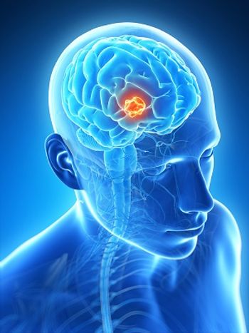
This case illustrates important endocrine sequelae that can be seen in patients with brain tumors, the most common malignancy in childhood. Craniopharyngiomas, histologically benign tumors, account for about 10% of all pediatric brain tumors.[1] They occur in the center of the brain near vital structures such as the optic nerve, hypothalamus, or pituitary and tend to adhere to surrounding delicate brain tissue. Treatment includes surgical resection, radiation therapy, or both[2,3] and results in an overall survival of 80% to 90%.[4,5]
FIGURE 1
Pituitary hormones and their target organs-ACTH, adrenocorticotropic hormone; GH, growth hormone; MSH, melanocyte-stimulating hormone; TSH, thyroid-stimulating hormone; FSH, follicle-stimulating hormone; LH, luteinizing hormone; ADH, antidiuretic hormone.
Damage to the pituitary and hypothalamus can occur from tumor extension or compression, during surgical resection or secondary to radiation therapy. The consequences of such damage are devastating. Figure 1 illustrates the many pituitary hormones and their diverse functions on target organs. These hormones promote body growth, protein synthesis, and fertility. They regulate glucose metabolism, cortisol/thyroid hormone production, and urine output.
The hypothalamus links the endocrine system with the neurologic system. It regulates body temperature, emotions, and energy balance. In adult survivors of craniopharyngioma, metabolic complications such as poor adrenal function, hypoglycemia, and antidiuretic hormone deficiency contribute to excessive mortality.[6] Patients may require maintenance replacement of one or more of the pituitary hormones. Survivors may require “stress dosing” of corticosteroids, such as prednisone. They are instructed to take the medication when experiencing stressful life events such as illness or accidents.
Less understood neuroendocrine functions such as hunger and satiety are also altered after damage to the pituitary or hypothalamus. Hypothalamic obesity occurs in half of craniopharyngioma survivors and is characterized by dysregulation of energy balance, resulting in excessive food intake and decreased caloric expenditure. Patients develop hyperinsulinism and exhibit uncontrolled, compulsive eating, which results in obesity. Many factors correlate with uncontrolled weight gain in craniopharyngioma survivors, including the tumor size and location, obesity before diagnosis, the need for shunt placement, the use of growth hormone or vasopressin, and the prolonged use of corticosteroids.
In addition, survivors who have hypothalamic damage from radiation doses in excess of 50 Gy have a higher risk of obesity.[7] This obesity predisposes patients to cardiovascular and cerebrovascular mortality,[8,9] particularly cerebrovascular accidents, transient ischemic events, and myocardial infarctions. One small series estimated that the overall risk of one of these events was 22% in craniopharyngioma survivors.[9]
Nursing Management
Adult survivors of childhood brain tumors present complex problems requiring careful evaluation and management. They are at high risk for metabolic complications and recurrent disease. Table 1 summarizes a risk-based assessment for survivors. First consideration must be given to evaluating continued tumor remission status.
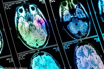
Magnetic resonance imaging of the brain is the most sensitive tool for detecting tumor progression. However, the community standard of care is to perform such a study in long-term survivors only if there has been a change in performance. Therefore, nurses should be diligent in helping survivors identify any deterioration in health or mental status.
Other items listed in Table 1 focus on monitoring metabolic abnormalities that can result from damage to the pituitary and hypothalamus and changes in normal hormonal production. Anthropometrics and measurement of body composition should be tracked from the time of diagnosis. Graphing weight and body mass index is helpful in demonstrating changes to patients over time. Nurses need to be alert to development of obesity.[10]
Behavioral and pharmacologic interventions have shown limited efficacy in moderating weight gain. Research suggests promotion of dietary restriction and exercise should be more aggressive and implemented early in therapy-ideally from the time of diagnosis.[1]
It is important to ascertain how survivors with insufficient adrenocorticosteroid production who take stress doses of steroids define “stress.” Are they including both minor infections and more significant events? Is their steroid medication in an accessible location?
Craniopharyngioma survivors experience many challenges and a great deal of anxiety about their vulnerability. Frank discussions lessen this anxiety. We have shown that nurses can help to create a positive listening environment in which survivors relate complete and honest information about their problems.[11,12] Discussions should include issues affecting access to care (eg, employment, insurance, and financial hardship). Compliance with medications should be frankly discussed. A detailed review of systems is needed to elicit symptoms of endocrinopathy. The results of laboratory data should be interpreted and discussed with survivors. Nurses can also facilitate informal communication between patients with similar risks to lessen their feelings of isolation. Finally, nurses can be advocates of community-based healthy lifestyle programs and encourage survivors to participate in a comprehensive program of weight management and exercise.
Table 1. Risk-based assessment for adult survivors of childhood brain tumors
Assessment of new neurologic symptoms and, if indicated, imaging to document survivor remains free of active tumor
Clinical assessments
Anthropometrics and body composition
Comprehensive health questionnaire
Detailed review of systems to elicit symptoms of endocrinopathy
Survivor knowledge of current endocrine status, health risks, and plans for care
Laboratory data
References:
References
1. Poretti A, Grotzer MA, Ribi K, et al: Outcome of craniopharyngioma in children: Long-term complications and quality of life. Dev Med Child Neurol 46(4):220â229, 2004.
2. Karavitaki N, Cudlip S, Adams CB, et al: Craniopharyngiomas. Endocr Rev 27(4):371â397, 2006.
3. Garre ML, Cama A: Craniopharyngioma: Modern concepts in pathogenesis and treatment. Curr Opin Pediatr 19(4):471â479, 2007.
4. Morris EB, Gajjar A, Okuma JO, et al: Survival and late mortality in long-term survivors of pediatric CNS tumors. J Clin Oncol 25(12):1532â1538, 2007.
5. Merchant TE, Kiehna EN, Sanford, RA, et al: Craniopharyngioma: The St. Jude Children’s Research Hospital experience 1984-2001. Int J Radiat Oncol Biol Phys 53(3):533â542, 2002.
6. Stripp DCH, Maity A, Janss AJ, et al: Surgery with or without radiation therapy in the management of craniopharyngiomas in children and young adults. Int J Radiat Oncol Biol Phys 58(3):714â720, 2004.
7. Lustig RH, Post SR, Srivannaboon K, et al: Risk factors for the development of obesity in children surviving brain tumors. J Clin Endocrinol Metab 88(2):611â616, 2003.
8. Daousi C, Dunn AJ, Foy PM, et al: Endocrine and neuroanatomic features associated with weight gain and obesity in adult patients with hypothalamic damage. Am J Med 118(1):45â50, 2005.
9. Pereira AM, Schmid EM, Schutte PJ, et al: High prevalence of long-term cardiovascular, neurological and psychosocial morbidity after treatment for craniopharyngioma. Clin Endocrinol 62(2):197â204, 2005.
10. Gleeson HK, Shalet SM: The impact of cancer therapy on the endocrine system in survivors of childhood brain tumours. Endocr Relat Cancer 11(4):589â602, 2004.
11. Crom DB, Hinds PS, Gattuso JS, et al: Creating the basis for a breast health program for female survivors of Hodgkin disease using a participatory research approach. Oncol Nurs Forum 32(6):1131â1141, 2005.
12. Crom DB, Tyc VL, Rai SN, et al: Retention of survivors of acute lymphoblastic leukemia in a longitudinal study of bone mineral density. J Child Health Care 10(4):337â50, 2006.
Articles in this issue
over 17 years ago
Review of "Physical Late Effects in Adult Cancer Survivors"over 17 years ago
Management of a Patient With Inflammatory Breast Cancerover 17 years ago
Cancer Care for Now … and Laterover 17 years ago
Diarrhea in Cancer Patientsover 17 years ago
Drug Essentials Levoleucovorin, a Cytoprotectantover 17 years ago
Physical Late Effects in Adult Cancer Survivorsover 17 years ago
Commentary (Cardonick): Care of the Pregnant Patient With Cancerover 17 years ago
Care of the Pregnant Patient With Canceralmost 18 years ago
Quality of Life in Myelodysplastic SyndromesNewsletter
Stay up to date on recent advances in the multidisciplinary approach to cancer.


